Division of Abdominal Imaging, Joint Department of Medical Imaging, University Health Network - Mount Sinai Hospital - Women's College Hospital, University of Toronto, Toronto, ON, Canada.
Insights Imaging. 2014 Feb;5(1):53-65. doi: 10.1007/s13244-013-0294-0. Epub 2013 Nov 29.
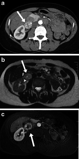
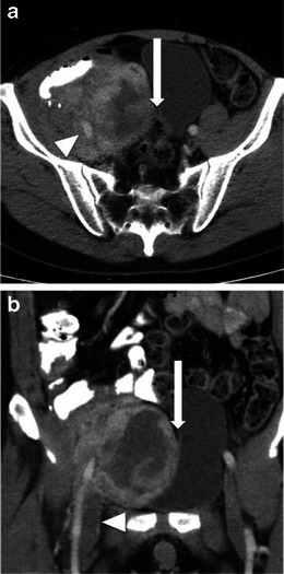
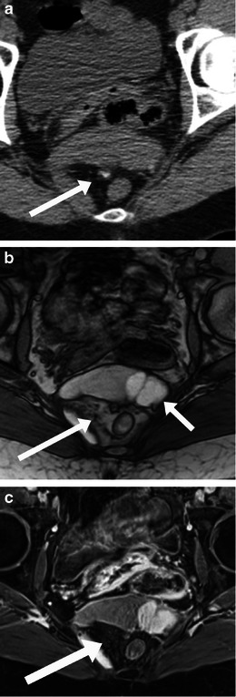
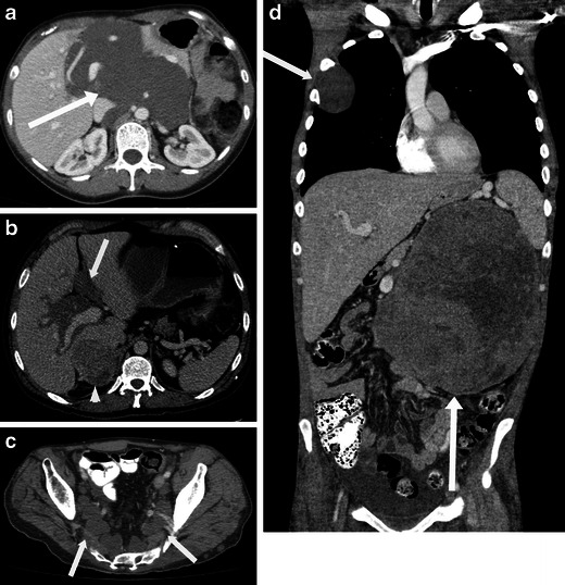
Primary retroperitoneal masses are a rare but important group of neoplasms. Cross-sectional imaging has revolutionised the investigation of patients with retroperitoneal neoplasms. Both computed tomography (CT) and magnetic resonance imaging (MRI) can contribute to tumour diagnosis, though histological confirmation is often required because of the considerable overlap of imaging features. Cross-sectional imaging is key to the pre-operative staging and planning of retroperitoneal masses, though ultrasound may also help in certain instances. Imaging also helps to select and guide the site to biopsy from these usually large and heterogeneous neoplasms. This article aims to review many of the primary retroperitoneal neoplasms that may be encountered by the radiologist.
原发性腹膜后肿块是一组罕见但重要的肿瘤。横断面成像技术已经彻底改变了腹膜后肿瘤患者的检查方式。计算机断层扫描(CT)和磁共振成像(MRI)都可以帮助诊断肿瘤,尽管由于影像学特征的显著重叠,通常需要组织学确认。横断面成像对于腹膜后肿块的术前分期和计划至关重要,但在某些情况下,超声也可能有所帮助。影像学还有助于从这些通常较大且异质性的肿瘤中选择并指导活检部位。本文旨在回顾放射科医生可能遇到的许多原发性腹膜后肿瘤。