Pallavaram Srivatsan, DʼHaese Pierre-François, Lake Wendell, Konrad Peter E, Dawant Benoit M, Neimat Joseph S
*Department of Electrical Engineering and Computer Science, Vanderbilt University, Nashville, Tennessee; ‡Department of Neurosurgery, Vanderbilt University Medical Center, Nashville, Tennessee.
Neurosurgery. 2015 Jun;76(6):756-65. doi: 10.1227/NEU.0000000000000714.
Finding the optimal location for the implantation of the electrode in deep brain stimulation (DBS) surgery is crucial for maximizing the therapeutic benefit to the patient. Such targeting is challenging for several reasons, including anatomic variability between patients as well as the lack of consensus about the location of the optimal target.
To compare the performance of popular manual targeting methods against a fully automatic nonrigid image registration-based approach.
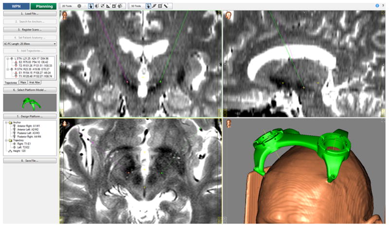
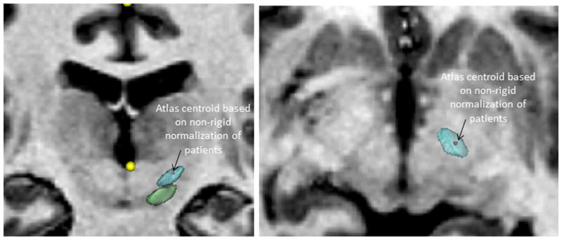
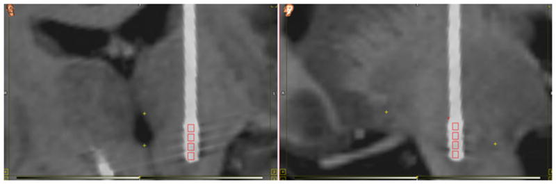
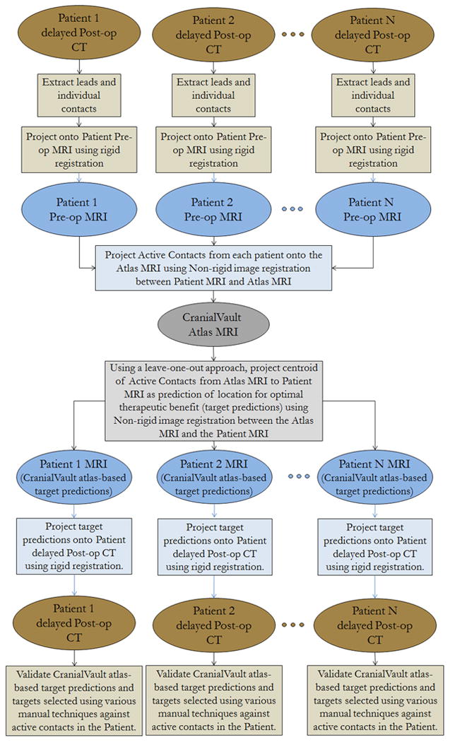
In 71 Parkinson disease subthalamic nucleus (STN)-DBS implantations, an experienced functional neurosurgeon selected the target manually using 3 different approaches: indirect targeting using standard stereotactic coordinates, direct targeting based on the patient magnetic resonance imaging, and indirect targeting relative to the red nucleus. Targets were also automatically predicted by using a leave-one-out approach to populate the CranialVault atlas with the use of nonrigid image registration. The different targeting methods were compared against the location of the final active contact, determined through iterative clinical programming in each individual patient.
Targeting by using standard stereotactic coordinates corresponding to the center of the motor territory of the STN had the largest targeting error (3.69 mm), followed by direct targeting (3.44 mm), average stereotactic coordinates of active contacts from this study (3.02 mm), red nucleus-based targeting (2.75 mm), and nonrigid image registration-based automatic predictions using the CranialVault atlas (2.70 mm). The CranialVault atlas method had statistically smaller variance than all manual approaches.
Fully automatic targeting based on nonrigid image registration with the use of the CranialVault atlas is as accurate and more precise than popular manual methods for STN-DBS.
在脑深部电刺激(DBS)手术中,找到电极植入的最佳位置对于使患者获得最大治疗益处至关重要。由于多种原因,这种靶点定位具有挑战性,包括患者之间的解剖变异以及关于最佳靶点位置缺乏共识。
比较常用手动靶点定位方法与基于全自动非刚性图像配准方法的性能。
在71例帕金森病丘脑底核(STN)-DBS植入手术中,一位经验丰富的功能神经外科医生使用3种不同方法手动选择靶点:使用标准立体定向坐标进行间接靶点定位、基于患者磁共振成像进行直接靶点定位以及相对于红核进行间接靶点定位。还通过使用留一法,利用非刚性图像配准将颅骨图谱应用于每个患者来自动预测靶点。将不同的靶点定位方法与通过对每个患者进行迭代临床编程确定的最终有效触点位置进行比较。
使用对应于STN运动区中心的标准立体定向坐标进行靶点定位的误差最大(3.69毫米),其次是直接靶点定位(3.44毫米)、本研究中有效触点的平均立体定向坐标(3.02毫米)、基于红核的靶点定位(2.75毫米)以及使用颅骨图谱的基于非刚性图像配准的自动预测(2.70毫米)。颅骨图谱方法的方差在统计学上小于所有手动方法。
对于STN-DBS,基于使用颅骨图谱的非刚性图像配准的全自动靶点定位与常用手动方法一样准确且更精确。