Colvin Michael T, Silvers Robert, Frohm Birgitta, Su Yongchao, Linse Sara, Griffin Robert G
†Department of Chemistry and Francis Bitter Magnet Laboratory, Massachusetts Institute of Technology, Cambridge, Massachusetts 02139, United States.
‡Department of Biochemistry and Structural Biology, Lund University, SE22100 Lund, Sweden.
J Am Chem Soc. 2015 Jun 17;137(23):7509-18. doi: 10.1021/jacs.5b03997. Epub 2015 Jun 4.
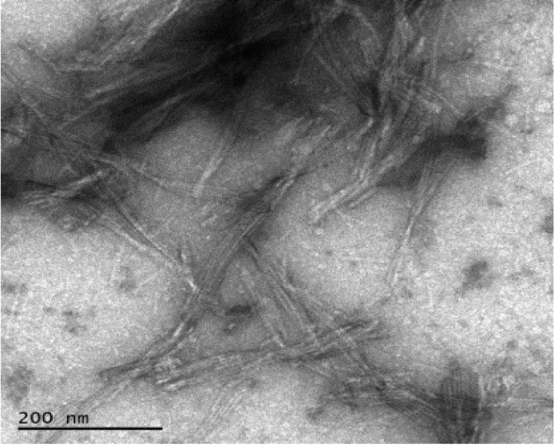
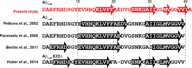
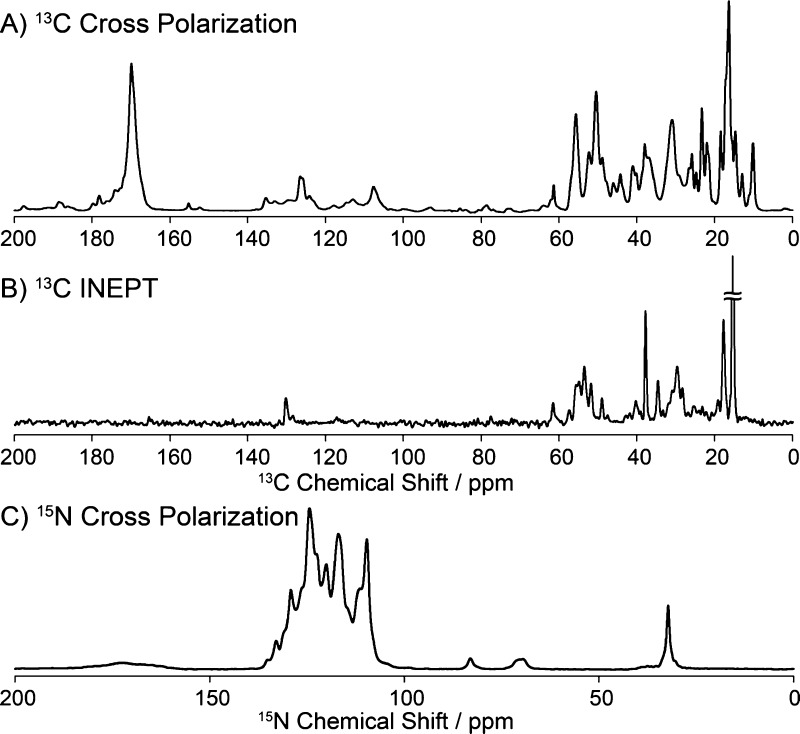
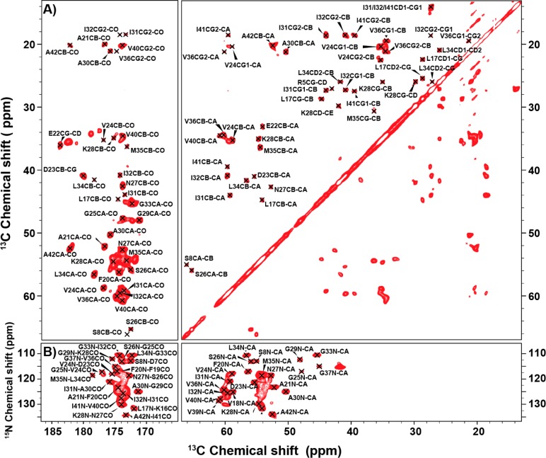
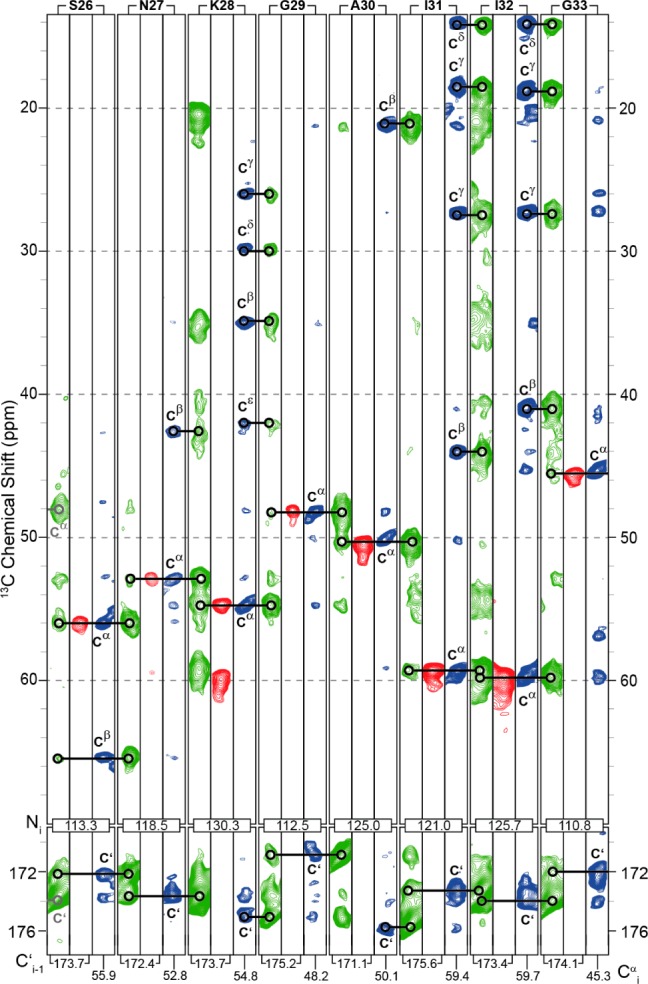
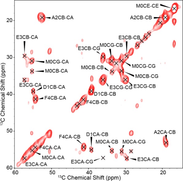
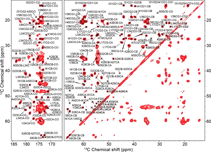
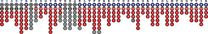
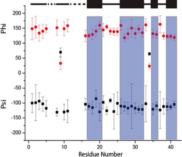
The presence of amyloid plaques composed of amyloid beta (Aβ) fibrils is a hallmark of Alzheimer's disease (AD). The Aβ peptide is present as several length variants with two common alloforms consisting of 40 and 42 amino acids, denoted Aβ1-40 and Aβ1-42, respectively. While there have been numerous reports that structurally characterize fibrils of Aβ1-40, very little is known about the structure of amyloid fibrils of Aβ1-42, which are considered the more toxic alloform involved in AD. We have prepared isotopically (13)C/(15)N labeled AβM01-42 fibrils in vitro from recombinant protein and examined their (13)C-(13)C and (13)C-(15)N magic angle spinning (MAS) NMR spectra. In contrast to several other studies of Aβ fibrils, we observe spectra with excellent resolution and a single set of chemical shifts, suggesting the presence of a single fibril morphology. We report the initial structural characterization of AβM01-42 fibrils utilizing (13)C and (15)N shift assignments of 38 of the 43 residues, including the backbone and side chains, obtained through a series of cross-polarization based 2D and 3D (13)C-(13)C, (13)C-(15)N MAS NMR experiments for rigid residues along with J-based 2D TOBSY experiments for dynamic residues. We find that the first ∼5 residues are dynamic and most efficiently detected in a J-based TOBSY spectrum. In contrast, residues 16-42 are easily observed in cross-polarization experiments and most likely form the amyloid core. Calculation of ψ and φ dihedral angles from the chemical shift assignments indicate that 4 β-strands are present in the fibril's secondary structure.
由β-淀粉样蛋白(Aβ)原纤维组成的淀粉样斑块的存在是阿尔茨海默病(AD)的一个标志。Aβ肽以几种长度变体形式存在,有两种常见的异构体,分别由40个和42个氨基酸组成,分别表示为Aβ1-40和Aβ1-42。虽然有许多关于Aβ1-40原纤维结构特征的报道,但对于Aβ1-42淀粉样原纤维的结构知之甚少,Aβ1-42被认为是参与AD的毒性更强的异构体。我们已经从重组蛋白体外制备了同位素(13)C/(15)N标记的AβM01-42原纤维,并检查了它们的(13)C-(13)C和(13)C-(15)N魔角旋转(MAS)NMR光谱。与其他几项关于Aβ原纤维的研究不同,我们观察到具有出色分辨率和单组化学位移的光谱,表明存在单一的原纤维形态。我们报告了利用通过一系列基于交叉极化的二维和三维(13)C-(13)C、(13)C-(15)N MAS NMR实验对刚性残基以及基于J的二维TOBSY实验对动态残基获得的43个残基中的38个残基(包括主链和侧链)的(13)C和(15)N位移归属对AβM01-42原纤维进行的初步结构表征。我们发现前约5个残基是动态的,并且在基于J的TOBSY光谱中最有效地被检测到。相比之下,残基16-42在交叉极化实验中很容易观察到,并且很可能形成淀粉样核心。根据化学位移归属计算的ψ和φ二面角表明,原纤维的二级结构中存在4条β链。