Department of Chemistry and Francis Bitter Magnet Laboratory, Massachusetts Institute of Technology , Cambridge, Massachusetts 02139, United States.
Bruker BioSpin , 15 Fortune Drive, Billerica, Massachusetts 01821, United States.
J Am Chem Soc. 2016 Aug 3;138(30):9663-74. doi: 10.1021/jacs.6b05129. Epub 2016 Jul 14.
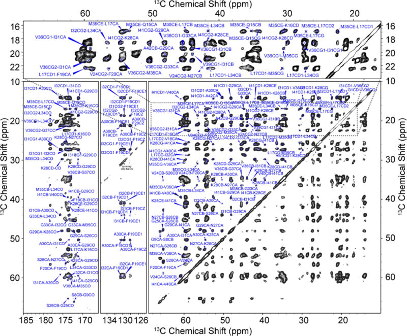
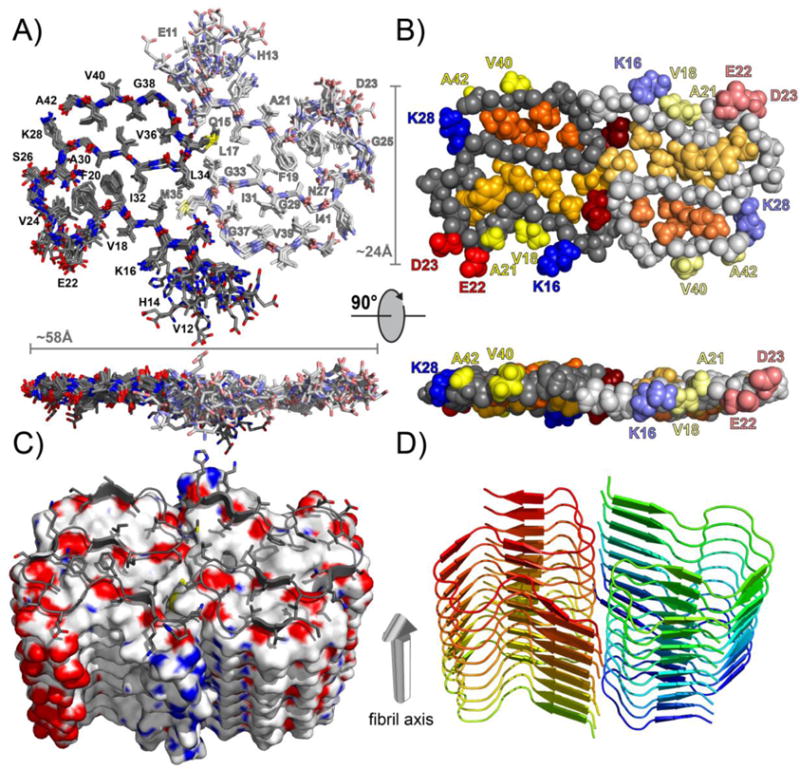
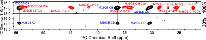
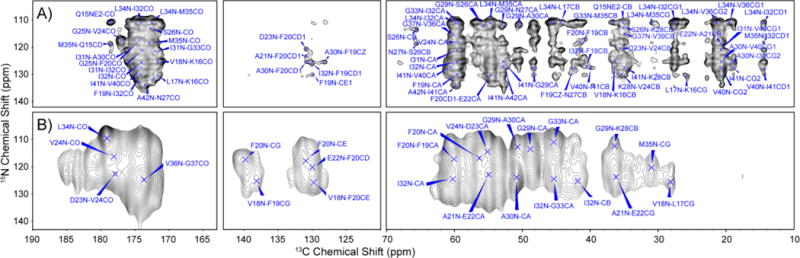
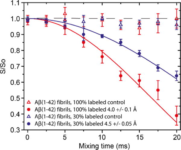
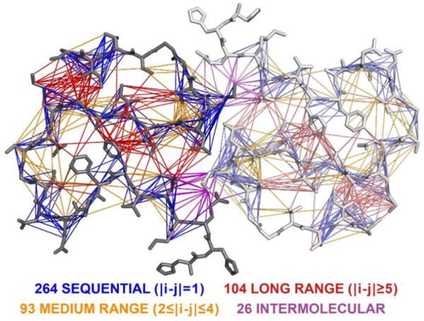
Amyloid-β (Aβ) is a 39-42 residue protein produced by the cleavage of the amyloid precursor protein (APP), which subsequently aggregates to form cross-β amyloid fibrils that are a hallmark of Alzheimer's disease (AD). The most prominent forms of Aβ are Aβ1-40 and Aβ1-42, which differ by two amino acids (I and A) at the C-terminus. However, Aβ42 is more neurotoxic and essential to the etiology of AD. Here, we present an atomic resolution structure of a monomorphic form of AβM01-42 amyloid fibrils derived from over 500 (13)C-(13)C, (13)C-(15)N distance and backbone angle structural constraints obtained from high field magic angle spinning NMR spectra. The structure (PDB ID: 5KK3 ) shows that the fibril core consists of a dimer of Aβ42 molecules, each containing four β-strands in a S-shaped amyloid fold, and arranged in a manner that generates two hydrophobic cores that are capped at the end of the chain by a salt bridge. The outer surface of the monomers presents hydrophilic side chains to the solvent. The interface between the monomers of the dimer shows clear contacts between M35 of one molecule and L17 and Q15 of the second. Intermolecular (13)C-(15)N constraints demonstrate that the amyloid fibrils are parallel in register. The RMSD of the backbone structure (Q15-A42) is 0.71 ± 0.12 Å and of all heavy atoms is 1.07 ± 0.08 Å. The structure provides a point of departure for the design of drugs that bind to the fibril surface and therefore interfere with secondary nucleation and for other therapeutic approaches to mitigate Aβ42 aggregation.
淀粉样蛋白-β(Aβ)是一种由淀粉样前体蛋白(APP)切割产生的 39-42 个残基蛋白,随后聚集形成交叉-β淀粉样纤维,这是阿尔茨海默病(AD)的标志。Aβ的最主要形式是 Aβ1-40 和 Aβ1-42,它们在 C 末端相差两个氨基酸(I 和 A)。然而,Aβ42 更具神经毒性,对 AD 的病因至关重要。在这里,我们展示了一种来自超过 500 个(13)C-(13)C、(13)C-(15)N 距离和从高场魔角旋转 NMR 光谱获得的 backbone 角结构约束的单形 AβM01-42 淀粉样纤维的原子分辨率结构。该结构(PDB ID:5KK3)显示纤维核心由 Aβ42 分子的二聚体组成,每个分子包含四个β-折叠的 S 形淀粉样纤维,排列方式产生两个疏水核心,在链的末端由盐桥封闭。单体的外表面向溶剂呈现亲水性侧链。二聚体单体之间的界面显示出一个分子的 M35 与第二个分子的 L17 和 Q15 之间的清晰接触。分子间(13)C-(15)N 约束证明淀粉样纤维是平行的。backbone 结构(Q15-A42)的 RMSD 为 0.71 ± 0.12 Å,所有重原子的 RMSD 为 1.07 ± 0.08 Å。该结构为设计结合纤维表面的药物提供了一个起点,这些药物可以干扰二级成核,以及为减轻 Aβ42 聚集的其他治疗方法提供了一个起点。