Pedoia V, Lansdown D A, Zaid M, McCulloch C E, Souza R, Ma C B, Li X
Department of Radiology and Biomedical Imaging, University of California, San Francisco, USA.
Department of Orthopaedic Surgery, University of California, San Francisco, USA.
Osteoarthritis Cartilage. 2015 Oct;23(10):1695-703. doi: 10.1016/j.joca.2015.05.027. Epub 2015 Jun 5.
The aim of this study is to develop a novel 3D magnetic resonance imaging (MRI)-based Statistical Shape Modeling (SSM) and apply it in knee MRIs in order to extract and compare relevant shapes of the tibia and femur in patients with and without acute Anterior cruciate ligament (ACL) injuries.
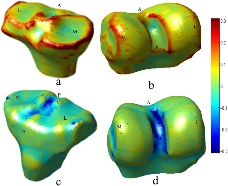
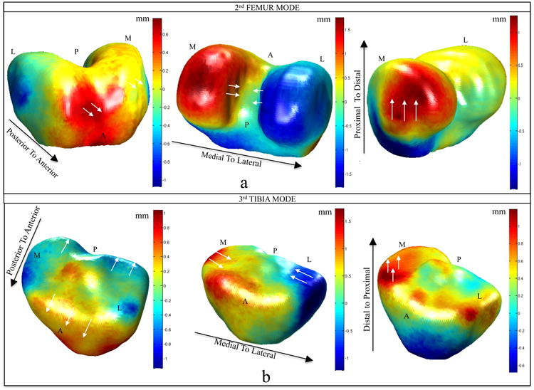
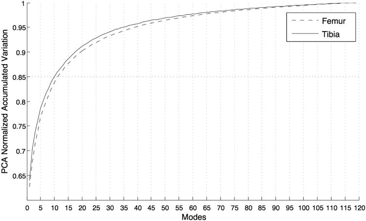
Bilateral MR images were acquired and analyzed for 50 patients with acute ACL injuries and for 19 control subjects. A shape model was extracted for the tibia and femur using an SSM algorithm based on a set of matched landmarks that are computed in a fully automatic manner.
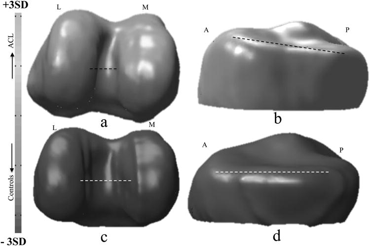
Shape differences were detected between the knees in the ACL-injury group and control group, suggesting a common shape feature that may predispose these knees to injury. Some of the detected shape features that discriminate between injured and control knees are related to intercondylar width and posterior tibia slope, features that have been suggested in previous studies as ACL morphological risk factors. However, shape modeling has the great potential to quantify these characteristics with a comprehensive description of the surfaces describing complex 3D deformation that cannot be represented with simple geometric indexes.
3D MRI-based bone shape quantification has the ability to identify specific anatomic risk factors for ACL injury. A better understanding of the role in bony shape on ligamentous injuries could help in the identification of subjects with an increased risk for an ACL tear and to develop targeted prevention strategies, including education and training.
本研究旨在开发一种基于三维磁共振成像(MRI)的新型统计形状建模(SSM),并将其应用于膝关节MRI,以提取和比较急性前交叉韧带(ACL)损伤患者和未损伤患者胫骨和股骨的相关形状。
对50例急性ACL损伤患者和19例对照受试者进行双侧MR图像采集和分析。使用基于一组以全自动方式计算的匹配地标点的SSM算法,为胫骨和股骨提取形状模型。
在ACL损伤组和对照组的膝关节之间检测到形状差异,表明存在一种可能使这些膝关节易受损伤的共同形状特征。一些在受伤膝关节和对照膝关节之间区分的检测到的形状特征与髁间宽度和胫骨后倾有关,这些特征在先前的研究中被认为是ACL形态学危险因素。然而,形状建模具有很大的潜力,可以通过对描述复杂三维变形的表面进行全面描述来量化这些特征,而这些变形无法用简单的几何指标表示。
基于三维MRI的骨形状量化能够识别ACL损伤的特定解剖学危险因素。更好地理解骨形状在韧带损伤中的作用有助于识别ACL撕裂风险增加的受试者,并制定有针对性的预防策略,包括教育和培训。