Liu Xuming, Yan Zhihan, Wang Tingyu, Yang Xiaokai, Feng Feng, Fan Luping, Jiang Jian
Department of Radiology, The Third Clinical Institute Affiliated to Wenzhou Medical University, Wenzhou, People's Republic of China.
Department of Radiology, The 2nd Affiliated Hospital of Wenzhou Medical University, Wenzhou, People's Republic of China.
Neuropsychiatr Dis Treat. 2015 May 26;11:1279-89. doi: 10.2147/NDT.S84204. eCollection 2015.
The aim of this study was to use functional magnetic resonance imaging (fMRI) technique to explore the resting-state functional connectivity (rsFC) differences of the bilaterial cerebellum posterior lobe (CPL) after normal sleep (NS) and after sleep deprivation (SD).
A total of 16 healthy subjects (eight males, eight females) underwent an fMRI scan twice at random: once following NS and the other following 24 hours' SD, with an interval of 1 month between the two scans. The fMRI scanning included resting state and acupuncture stimulation. The special activated regions located during the acupuncture stimulation were selected as regions of interest for rsFC analysis.
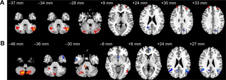
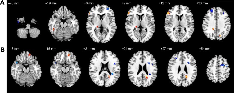
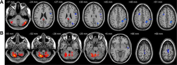
Bilateral CPLs were positively activated by acupuncture stimulation. In the NS group, the left CPL showed rsFC with the bilateral CPL, bilateral frontal lobe (BFL), left precuneus and right inferior parietal lobule, while the right CPL showed rsFC with the bilateral temporal lobe, right cerebellum anterior lobe, right CPL, left frontal lobe, left anterior cingulate, right posterior cingulate, and bilateral inferior parietal lobule. In the SD group, the left CPL showed rsFC with the left posterior cingulate gyrus bilateral CPL, left precuneus, left precentral gyrus, BFL, and the left parietal lobe, while the right CPL showed rsFC with bilateral cerebellum anterior lobe, bilateral CPL, left frontal lobe and left temporal lobe. Compared with the NS group, the left CPL had increased rsFC in the SD group with the right inferior frontal gyrus, right fusiform gyrus, right cingulate gyrus, right thalamus, and bilateral precuneus, and decreased rsFC with the BFL, while the right CPL had increased rsFC with the left superior frontal gyrus and decreased rsFC with the left precentral gyrus, right superior temporal gyrus, and the BFL.
Bilateral CPL are possibly involved in acupuncture stimulation in different manners, and the right CPL showed more rsFC impairment.
本研究旨在运用功能磁共振成像(fMRI)技术,探究正常睡眠(NS)和睡眠剥夺(SD)后双侧小脑后叶(CPL)的静息态功能连接(rsFC)差异。
共有16名健康受试者(8名男性,8名女性)随机接受两次fMRI扫描:一次在正常睡眠后,另一次在24小时睡眠剥夺后,两次扫描间隔1个月。fMRI扫描包括静息状态和针刺刺激。将针刺刺激时定位的特殊激活区域选为rsFC分析的感兴趣区域。
双侧CPL在针刺刺激下呈阳性激活。在正常睡眠组中,左侧CPL与双侧CPL、双侧额叶(BFL)、左侧楔前叶和右侧顶下小叶显示rsFC,而右侧CPL与双侧颞叶、右侧小脑前叶、右侧CPL、左侧额叶、左侧前扣带回、右侧后扣带回和双侧顶下小叶显示rsFC。在睡眠剥夺组中,左侧CPL与左侧后扣带回、双侧CPL、左侧楔前叶、左侧中央前回、BFL和左侧顶叶显示rsFC,而右侧CPL与双侧小脑前叶、双侧CPL、左侧额叶和左侧颞叶显示rsFC。与正常睡眠组相比,睡眠剥夺组中左侧CPL与右侧额下回、右侧梭状回、右侧扣带回、右侧丘脑和双侧楔前叶的rsFC增加,与BFL的rsFC减少,而右侧CPL与左侧额上回的rsFC增加,与左侧中央前回、右侧颞上回和BFL的rsFC减少。
双侧CPL可能以不同方式参与针刺刺激,且右侧CPL显示出更多的rsFC损伤。