Kondo Kosuke, Nemoto Masaaki, Masuda Hiroyuki, Okonogi Shinichi, Nomoto Jun, Harada Naoyuki, Sugo Nobuo, Miyazaki Chikao
Department of Neurosurgery (Omori), School of Medicine, Toho University.
Neurol Med Chir (Tokyo). 2015;55(7):592-8. doi: 10.2176/nmc.oa.2014-0436. Epub 2015 Jun 29.
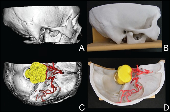
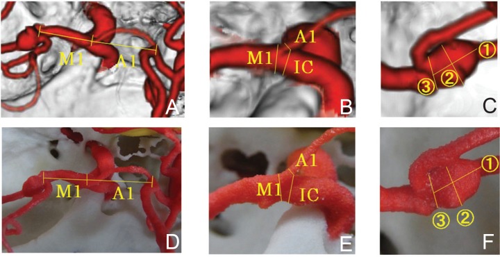
We prepared rapid prototyping models of heads with unruptured cerebral aneurysm based on image data of computed tomography angiography (CTA) using a three-dimensional (3D) printer. The objective of this study was to evaluate the anatomical reproducibility and accuracy of these models by comparison with the CTA images on a monitor. The subjects were 22 patients with unruptured cerebral aneurysm who underwent preoperative CTA. Reproducibility of the microsurgical anatomy of skull bone and arteries, the length and thickness of the main arteries, and the size of cerebral aneurysm were compared between the CTA image and rapid prototyping model. The microsurgical anatomy and arteries were favorably reproduced, apart from a few minute regions, in the rapid prototyping models. No significant difference was noted in the measured lengths of the main arteries between the CTA image and rapid prototyping model, but errors were noted in their thickness (p < 0.001). A significant difference was also noted in the longitudinal diameter of the cerebral aneurysm (p < 0.01). Regarding the CTA image as the gold standard, reproducibility of the microsurgical anatomy of skull bone and main arteries was favorable in the rapid prototyping models prepared using a 3D printer. It was concluded that these models are useful tools for neurosurgical simulation. The thickness of the main arteries and size of cerebral aneurysm should be comprehensively judged including other neuroimaging in consideration of errors.
我们使用三维(3D)打印机,基于计算机断层扫描血管造影(CTA)的图像数据,制备了患有未破裂脑动脉瘤的头部快速成型模型。本研究的目的是通过与监视器上的CTA图像进行比较,评估这些模型的解剖学再现性和准确性。研究对象为22例接受术前CTA检查的未破裂脑动脉瘤患者。比较了CTA图像和快速成型模型之间颅骨和动脉的显微外科解剖结构的再现性、主要动脉的长度和厚度以及脑动脉瘤的大小。在快速成型模型中,除了一些微小区域外,显微外科解剖结构和动脉均得到了良好的再现。CTA图像和快速成型模型之间主要动脉的测量长度没有显著差异,但厚度存在误差(p < 0.001)。脑动脉瘤的纵向直径也存在显著差异(p < 0.01)。以CTA图像作为金标准,使用3D打印机制备的快速成型模型中颅骨和主要动脉的显微外科解剖结构再现性良好。得出的结论是,这些模型是神经外科模拟的有用工具。考虑到误差,应结合其他神经影像学检查,综合判断主要动脉的厚度和脑动脉瘤的大小。