Faraj Moneer K, Hoz Samer S, Mohammad Amjad J
Department of Neurosurgery, College of Medicine, Neurosciences Hospital, University of Baghdad, Iraq.
Department of Neurosurgery, Neurosurgery Teaching Hospital, Baghdad, Iraq.
Surg Neurol Int. 2020 Nov 11;11:381. doi: 10.25259/SNI_361_2020. eCollection 2020.
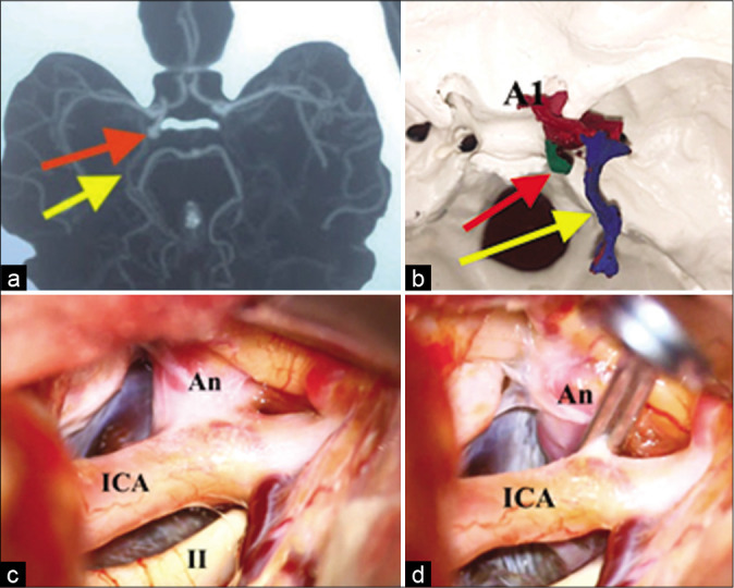
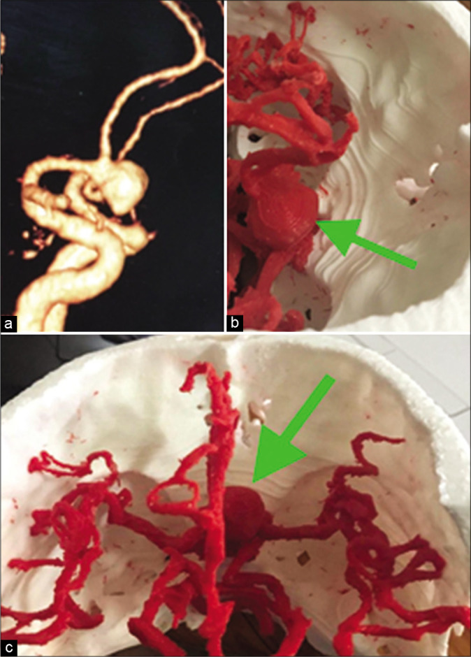
In the present study, we aim to develop simulation models based on computed tomography angiography images of intracranial aneurysms (IAs) and their parent vessels using three-dimensional (3D) printing technology. The study focuses on the value of these 3D models in presurgical planning and intraoperative navigation and ultimately their impact on patient outcomes. To the best of our knowledge, this is the first report of its kind from a war-torn country, like Iraq.
This is a prospective study of a series of 11, consecutively enrolled, patients suffering from IAs for the period between February and September 2019. The study represents a collaboration between the two major neurosurgical centers in Baghdad/Iraq; Neurosciences Teaching Hospital and Neurosurgery Teaching Hospital. We analyzed the data of eleven patients with IAs treated by microsurgical clipping. These data include patient demographics, clinical, surgical, and outcomes along with the data of the 3D-printed replica used in these surgeries. All cases were operated on by one surgeon.
Our study included 11 patients, with a total of 11 aneurysms clipped. The mean age was 44 ± 8, with a median of 42.5 and a range of 35-61 years. About 60% of our patients were female with a female-to-male ratio of 1:5. About 60% of the aneurysms were located at the anterior communicating artery (Acom) while the remaining 40% were equally distributed between the posterior communicating and internal carotid arteries bifurcation. The standard pterional approach was followed in 50% of cases, whereas the other 50% of patients were treated through the lateral supraorbital approach. About 90% ( = 9) of the patients had a Glasgow Outcome Scale (GOS) of 5 and 10% had a GOS of 4. The 3D-printed models successfully replicated the aneurysm size, location, and relation to the parent vessel with 100% accuracy and were used for intraoperative guidance. The average production time was 24-48 h and the production cost was 10-20 US dollars.
3D printing is a promising technology that is rapidly penetrating the field of neurosurgery. In particular, the use of 3D-printed patient-matched, anatomically accurate replicas of the cerebral vascular tree is valuable adjunct to the microsurgical clipping of IAs, and our study conclusions support this concept. However, both the feasibility and clinical utility of 3D printing remain the subject of much, ongoing investigations.
在本研究中,我们旨在利用三维(3D)打印技术,基于颅内动脉瘤(IA)及其供血血管的计算机断层血管造影图像开发仿真模型。本研究聚焦于这些3D模型在术前规划和术中导航中的价值,以及它们最终对患者预后的影响。据我们所知,这是来自伊拉克这样一个饱受战争蹂躏国家的同类首例报告。
这是一项前瞻性研究,对2019年2月至9月期间连续入选的11例IA患者进行了研究。该研究是伊拉克巴格达两家主要神经外科中心——神经科学教学医院和神经外科教学医院之间的合作项目。我们分析了11例接受显微手术夹闭治疗的IA患者的数据。这些数据包括患者的人口统计学信息、临床、手术及预后情况,以及这些手术中使用的3D打印复制品的数据。所有病例均由一名外科医生进行手术。
我们的研究纳入了11例患者,共夹闭了11个动脉瘤。平均年龄为44±8岁,中位数为42.5岁,范围为35 - 61岁。约60%的患者为女性,男女比例为1:5。约60%的动脉瘤位于前交通动脉(Acom),其余40%在后交通动脉和颈内动脉分叉处平均分布。50%的病例采用标准翼点入路,而其他50%的患者通过眶上外侧入路进行治疗。约90%(n = 9)的患者格拉斯哥预后量表(GOS)评分为5分,10%的患者GOS评分为4分。3D打印模型成功地以100%的准确率复制了动脉瘤的大小、位置及其与供血血管的关系,并用于术中指导。平均制作时间为24 - 48小时,制作成本为10 - 20美元。
3D打印是一项很有前景的技术,正在迅速渗透到神经外科领域。特别是,使用3D打印的与患者匹配的、解剖结构精确的脑血管树复制品,对于IA的显微手术夹闭是一种有价值的辅助手段,我们的研究结论支持这一概念。然而,3D打印的可行性和临床实用性仍是众多正在进行的研究的主题。