Klibanov Alexander L, Hossack John A
From the *Cardiovascular Division, Robert M. Berne Cardiovascular Research Center, School of Medicine, and †Department of Biomedical Engineering, University of Virginia, Charlottesville VA.
Invest Radiol. 2015 Sep;50(9):657-70. doi: 10.1097/RLI.0000000000000188.
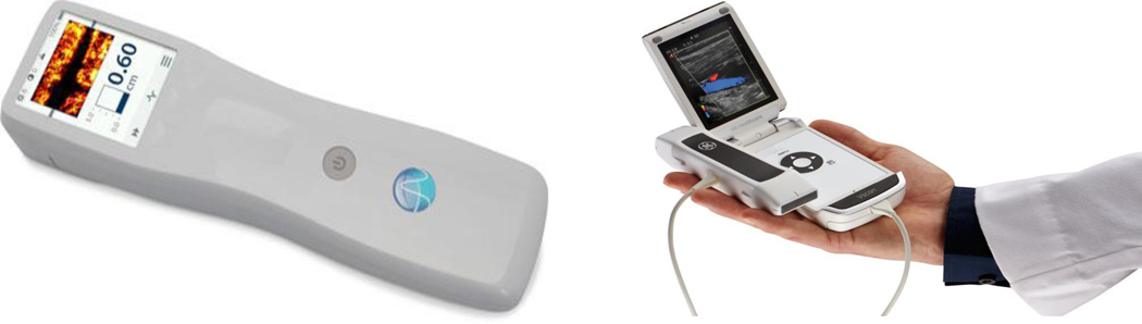
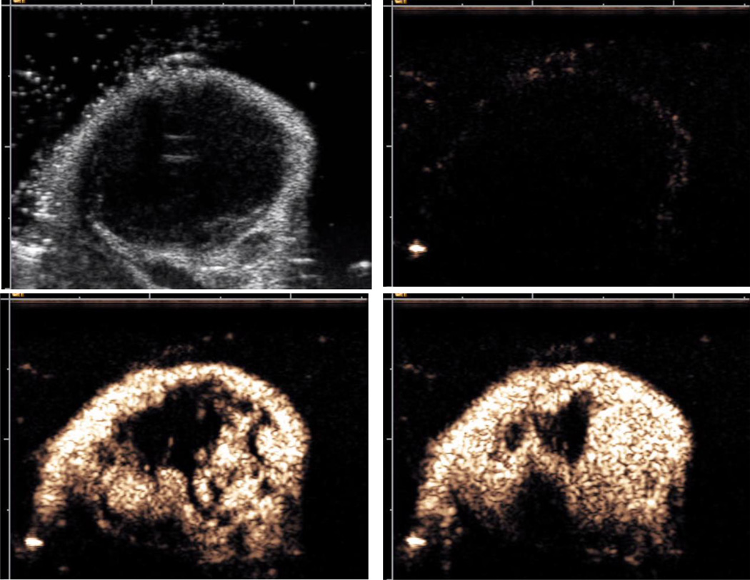
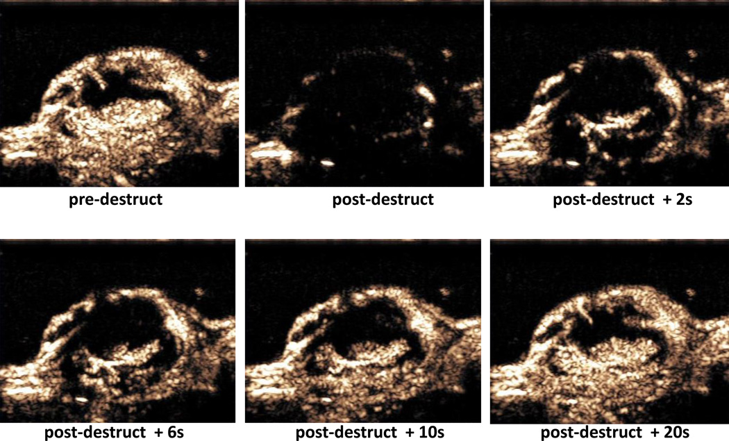
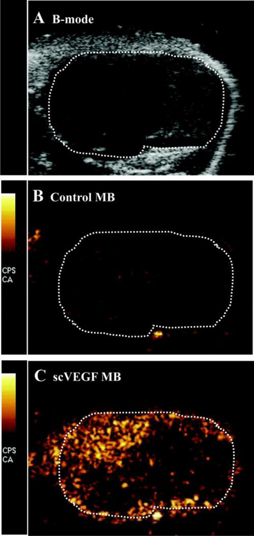
During the past decade, ultrasound has expanded medical imaging well beyond the "traditional" radiology setting: a combination of portability, low cost, and ease of use makes ultrasound imaging an indispensable tool for radiologists as well as for other medical professionals who need to obtain imaging diagnosis or guide a therapeutic intervention quickly and efficiently. Ultrasound combines excellent ability for deep penetration into soft tissues with very good spatial resolution, with only a few exceptions (ie, those involving overlying bone or gas). Real-time imaging (up to hundreds and thousands of frames per second) enables guidance of therapeutic procedures and biopsies; characterization of the mechanical properties of the tissues greatly aids with the accuracy of the procedures. The ability of ultrasound to deposit energy locally brings about the potential for localized intervention encompassing the following: tissue ablation, enhancing penetration through the natural barriers to drug delivery in the body and triggering drug release from carrier microparticles and nanoparticles. The use of microbubble contrast agents brings the ability to monitor and quantify tissue perfusion, and microbubble targeting with ligand-decorated microbubbles brings the ability to obtain molecular biomarker information, that is, ultrasound molecular imaging. Overall, ultrasound has become the most widely used imaging modality in modern medicine; it will continue to grow and expand.
在过去十年间,超声已将医学成像拓展至远远超出“传统”放射学范畴:便携性、低成本及易用性的结合,使超声成像成为放射科医生以及其他需要快速高效地获得影像诊断或指导治疗干预的医学专业人员不可或缺的工具。超声结合了深入软组织的卓越能力与良好的空间分辨率,仅有少数例外情况(即涉及覆盖骨骼或气体的情况)。实时成像(每秒可达数百乃至数千帧)能够指导治疗程序和活检;组织力学特性的表征极大地有助于提高这些程序的准确性。超声在局部沉积能量的能力带来了局部干预的潜力,包括:组织消融、增强药物穿过体内自然屏障的渗透以及触发药物从载体微泡和纳米颗粒中释放。微泡造影剂的使用带来了监测和量化组织灌注的能力,而用配体修饰的微泡进行微泡靶向则带来了获取分子生物标志物信息的能力,即超声分子成像。总体而言,超声已成为现代医学中使用最广泛的成像方式;它将继续发展和拓展。