Cheng Wei-Hung, Huang Kuo-Yang, Huang Po-Jung, Hsu Jo-Hsuan, Fang Yi-Kai, Chiu Cheng-Hsun, Tang Petrus
Graduate Institute of Biomedical Sciences, College of Medicine, Chang Gung University, Kweishan, Taoyuan, Taiwan.
Molecular Regulation and Bioinformatics Laboratory, Department of Parasitology, College of Medicine, Chang Gung University, Kweishan, Taoyuan, Taiwan.
Parasit Vectors. 2015 Jul 25;8:393. doi: 10.1186/s13071-015-1000-5.
Iron plays a pivotal role in the pathogenesis of Trichomonas vaginalis, the causative agent of highly prevalent human trichomoniasis. T. vaginalis resides in the vaginal region, where the iron concentration is constantly changing. Hence, T. vaginalis must adapt to variations in iron availability to establish and maintain an infection. The free radical signaling molecules reactive oxygen species (ROS) and reactive nitrogen species (RNS) have been proven to participate in iron deficiency in eukaryotes. However, little is known about the roles of these molecules in iron-deficient T. vaginalis.
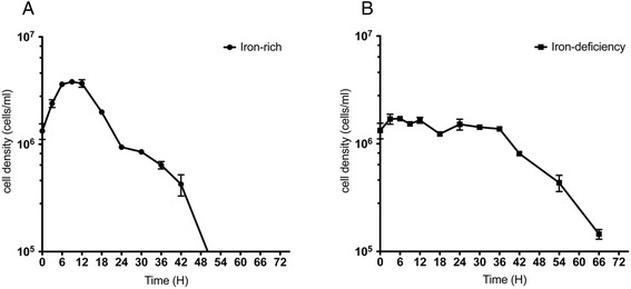
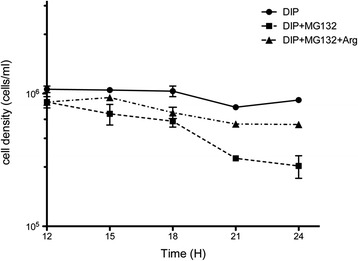
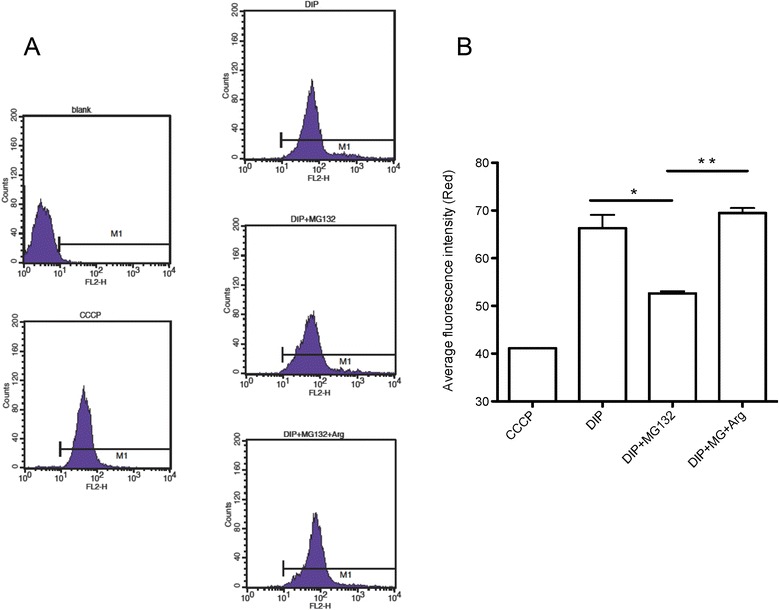
T. vaginalis cultured in iron-rich and -deficient conditions were collected for all experiments in this study. Next generation RNA sequencing was conducted to investigate the impact of iron on transcriptome of T. vaginalis. The cell viabilities were monitored after the trophozoites treated with the inhibitors of nitric oxide (NO) synthase (L-NG-monomethyl arginine, L-NMMA) and proteasome (MG132). Hydrogenosomal membrane potential was measured using JC-1 staining.
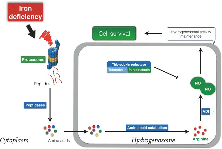
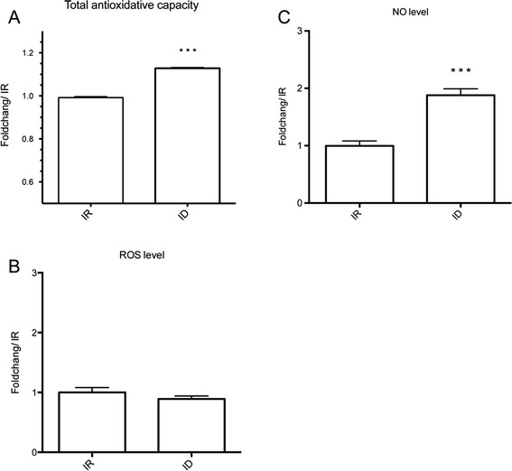
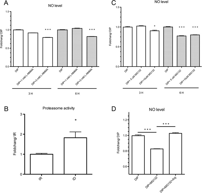
We demonstrated that NO rather than ROS accumulates in iron-deficient T. vaginalis. The level of NO was blocked by MG132 and L-NMMA, indicating that NO production is through a proteasome and arginine dependent pathway. We found that the inhibition of proteasome activity shortened the survival of iron-deficient cells compared with untreated iron-deficient cells. Surprisingly, the addition of arginine restored both NO level and the survival of proteasome-inhibited cells, suggesting that proteasome-derived NO is crucial for cell survival under iron-limited conditions. Additionally, NO maintains the hydrogenosomal membrane potential, a determinant for cell survival, emphasizing the cytoprotective effect of NO on iron-deficient T. vaginalis. Collectively, we determined that NO produced by the proteasome prolonged the survival of iron-deficient T. vaginalis via maintenance of the hydrogenosomal functions.
The findings in this study provide a novel role of NO in adaptation to iron-deficient stress in T. vaginalis and shed light on a potential therapeutic strategy for trichomoniasis.
铁在阴道毛滴虫的发病机制中起关键作用,阴道毛滴虫是人类滴虫病的病原体,该病高度流行。阴道毛滴虫寄居于阴道区域,此处铁浓度不断变化。因此,阴道毛滴虫必须适应铁可利用性的变化以建立并维持感染。自由基信号分子活性氧(ROS)和活性氮(RNS)已被证明参与真核生物的缺铁过程。然而,关于这些分子在缺铁的阴道毛滴虫中的作用知之甚少。
本研究中所有实验均收集在富铁和缺铁条件下培养的阴道毛滴虫。进行下一代RNA测序以研究铁对阴道毛滴虫转录组的影响。在用一氧化氮(NO)合酶抑制剂(L-NG-单甲基精氨酸,L-NMMA)和蛋白酶体抑制剂(MG132)处理滋养体后监测细胞活力。使用JC-1染色测量氢化酶体膜电位。
我们证明在缺铁的阴道毛滴虫中积累的是NO而非ROS。MG132和L-NMMA可阻断NO水平,表明NO的产生是通过蛋白酶体和精氨酸依赖性途径。我们发现与未处理的缺铁细胞相比,蛋白酶体活性的抑制缩短了缺铁细胞的存活时间。令人惊讶的是,添加精氨酸可恢复NO水平和蛋白酶体抑制细胞的存活率,表明蛋白酶体衍生的NO对于铁限制条件下的细胞存活至关重要。此外,NO维持氢化酶体膜电位,这是细胞存活的决定因素,强调了NO对缺铁阴道毛滴虫的细胞保护作用。总体而言,我们确定蛋白酶体产生的NO通过维持氢化酶体功能延长了缺铁阴道毛滴虫的存活时间。
本研究结果揭示了NO在阴道毛滴虫适应缺铁应激中的新作用,并为滴虫病的潜在治疗策略提供了线索。