Pulver Stefan R, Bayley Timothy G, Taylor Adam L, Berni Jimena, Bate Michael, Hedwig Berthold
School of Psychology and Neuroscience, University of St Andrews, St Andrews, United Kingdom; Janelia Research Campus, Howard Hughes Medical Institute, Ashburn, Virginia
Department of Zoology, University of Cambridge, Cambridge, United Kingdom; and.
J Neurophysiol. 2015 Nov;114(5):2564-77. doi: 10.1152/jn.00731.2015. Epub 2015 Aug 26.
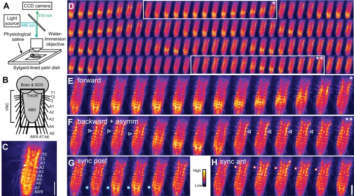
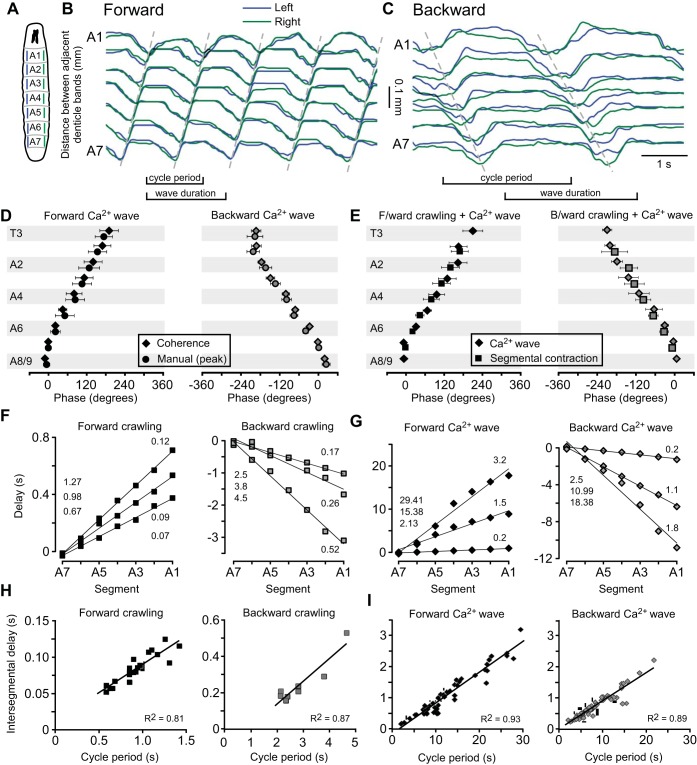
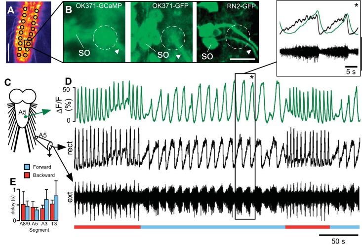
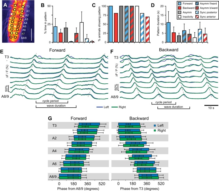
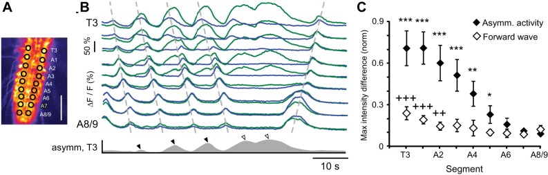
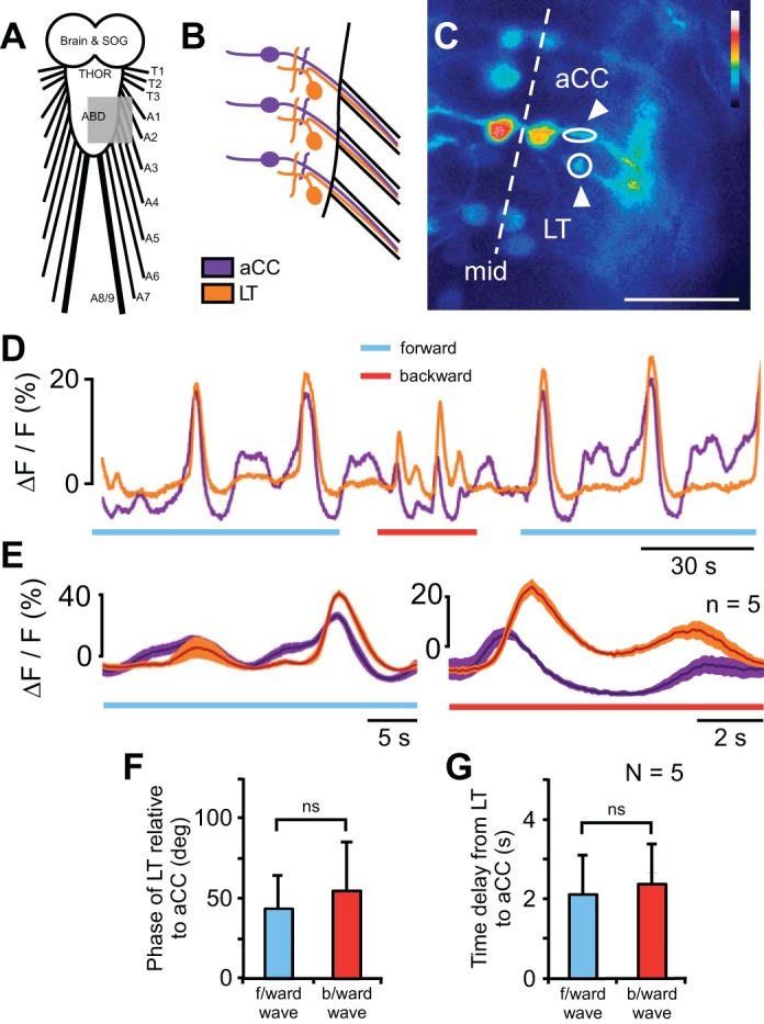
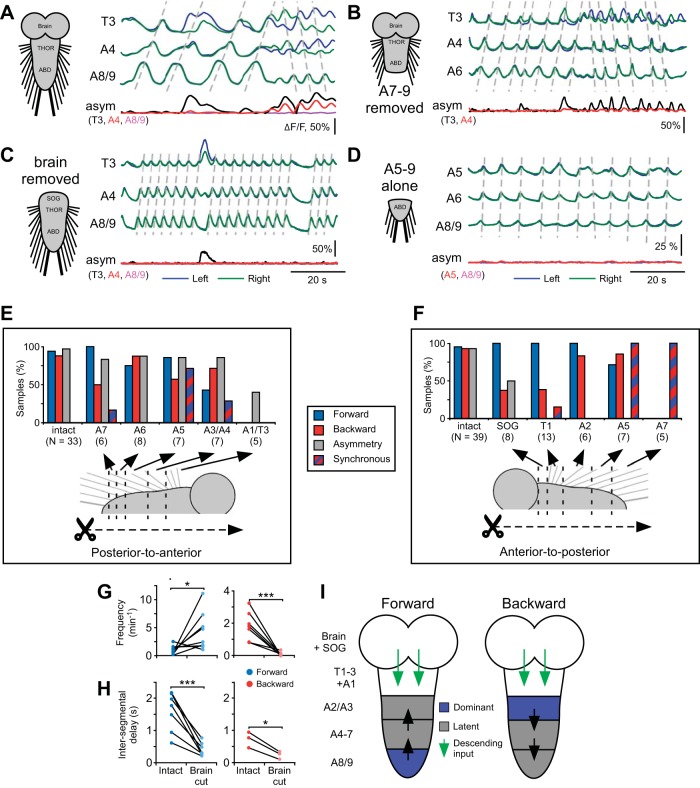
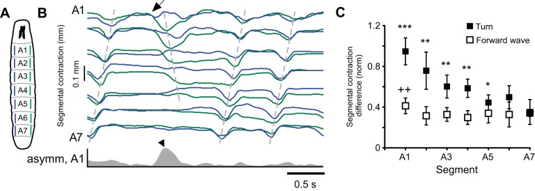
We have established a preparation in larval Drosophila to monitor fictive locomotion simultaneously across abdominal and thoracic segments of the isolated CNS with genetically encoded Ca(2+) indicators. The Ca(2+) signals closely followed spiking activity measured electrophysiologically in nerve roots. Three motor patterns are analyzed. Two comprise waves of Ca(2+) signals that progress along the longitudinal body axis in a posterior-to-anterior or anterior-to-posterior direction. These waves had statistically indistinguishable intersegmental phase delays compared with segmental contractions during forward and backward crawling behavior, despite being ∼10 times slower. During these waves, motor neurons of the dorsal longitudinal and transverse muscles were active in the same order as the muscle groups are recruited during crawling behavior. A third fictive motor pattern exhibits a left-right asymmetry across segments and bears similarities with turning behavior in intact larvae, occurring equally frequently and involving asymmetry in the same segments. Ablation of the segments in which forward and backward waves of Ca(2+) signals were normally initiated did not eliminate production of Ca(2+) waves. When the brain and subesophageal ganglion (SOG) were removed, the remaining ganglia retained the ability to produce both forward and backward waves of motor activity, although the speed and frequency of waves changed. Bilateral asymmetry of activity was reduced when the brain was removed and abolished when the SOG was removed. This work paves the way to studying the neural and genetic underpinnings of segmentally coordinated motor pattern generation in Drosophila with imaging techniques.
我们已经在果蝇幼虫中建立了一种制备方法,使用基因编码的Ca(2+)指示剂同时监测分离的中枢神经系统腹部和胸部各节段的虚拟运动。Ca(2+)信号紧密跟随在神经根中通过电生理学测量的动作电位活动。分析了三种运动模式。其中两种模式包括Ca(2+)信号波,它们沿身体纵轴以从后向前或从前向后的方向传播。尽管这些波的速度比向前和向后爬行行为期间的节段收缩慢约10倍,但与节段收缩相比,它们的节间相位延迟在统计学上没有差异。在这些波期间,背纵肌和横肌的运动神经元的激活顺序与爬行行为期间肌肉群的募集顺序相同。第三种虚拟运动模式在各节段之间表现出左右不对称,与完整幼虫的转向行为相似,出现频率相同且涉及相同节段的不对称。去除正常启动Ca(2+)信号向前和向后波的节段并不能消除Ca(2+)波的产生。当去除脑和咽下神经节(SOG)时,剩余的神经节仍保留产生运动活动向前和向后波的能力,尽管波的速度和频率发生了变化。去除脑时活动的双侧不对称性降低,去除SOG时则消除。这项工作为使用成像技术研究果蝇节段协调运动模式产生的神经和遗传基础铺平了道路。