Sevuk Utkan, Bahadir Mehmet Veysi, Altindag Rojhat, Baysal Erkan, Yaylak Baris, Ay Nurettin, Ayaz Firat, Demirtas Ertan
Department of Cardiovascular Surgery, Diyarbakir Gazi Yasargil Education and Research Hospital, Diyarbakir, Turkey.
Department of General Surgery, Dicle University, Diyarbakir, Turkey.
Ther Clin Risk Manag. 2015 Aug 20;11:1243-9. doi: 10.2147/TCRM.S89355. eCollection 2015.
To date, no validated biomarkers with high sensitivity and specificity have been established for diagnosis of pulmonary embolism (PE) in patients with deep venous thrombosis (DVT). There is a need to develop simple and reliable noninvasive tests that can accurately identify patients with PE, even in small hospitals or clinics. The aim of this study was to investigate the value of mean platelet volume (MPV) and platelet distribution width (PDW) for predicting occurrence of PE in patients with DVT.
Records of acute DVT patients were reviewed retrospectively. Group 1 consisted of 50 patients with acute DVT and group 2 consisted of 50 patients with acute DVT who developed PE during follow-up. The control group consisted of patients with uncomplicated primary varicose veins of the lower limbs. Venous peripheral blood samples for measurement of MPV, PDW, and platelet count were drawn on admission, before the treatment, and at the time of PE diagnosis.
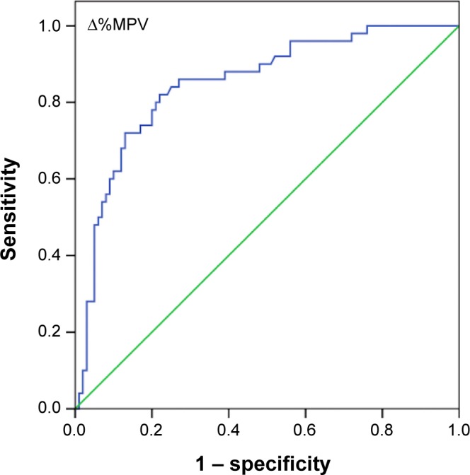
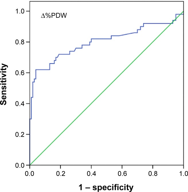
MPV and PDW levels at the time of PE diagnosis were higher in group 2 than group 1 (P<0.001 and P=0.026, respectively). Receiver operating characteristics analysis revealed that a 5.2% increase in admission PDW during follow-up provided 70% sensitivity and 82% specificity (area under the curve, 0.80), and a 6.6% increase in admission MPV during follow-up provided 74% sensitivity and 83% specificity (area under the curve, 0.84) for prediction of PE occurrence in patients with DVT. PDW and MPV levels at the time of PE diagnosis were found to be independent risk factors for the occurrence of PE in patients with DVT.
Serial measurements of MPV and PDW, and percent change in MPV and PDW appears to be a useful marker for predicting occurrence of acute PE in patients with a first episode of acute proximal DVT.
迄今为止,尚未建立用于诊断深静脉血栓形成(DVT)患者肺栓塞(PE)的具有高敏感性和特异性的经过验证的生物标志物。需要开发简单可靠的非侵入性检测方法,即使在小型医院或诊所也能准确识别PE患者。本研究的目的是探讨平均血小板体积(MPV)和血小板分布宽度(PDW)对预测DVT患者发生PE的价值。
回顾性分析急性DVT患者的记录。第1组由50例急性DVT患者组成,第2组由50例在随访期间发生PE的急性DVT患者组成。对照组由单纯性下肢原发性静脉曲张患者组成。在入院时、治疗前以及PE诊断时采集静脉外周血样本,用于测定MPV、PDW和血小板计数。
第2组PE诊断时的MPV和PDW水平高于第1组(分别为P<0.001和P=0.026)。受试者工作特征分析显示,随访期间入院时PDW增加5.2%,预测DVT患者发生PE的敏感性为70%,特异性为82%(曲线下面积为0.80);随访期间入院时MPV增加6.6%,预测DVT患者发生PE的敏感性为74%,特异性为83%(曲线下面积为0.84)。发现PE诊断时的PDW和MPV水平是DVT患者发生PE的独立危险因素。
对MPV和PDW进行系列测量,以及MPV和PDW的变化百分比似乎是预测首次发生急性近端DVT患者发生急性PE的有用标志物。