Nassar Zeyad D, Hill Michelle M, Parton Robert G, Francois Mathias, Parat Marie-Odile
The University of Queensland, School of Pharmacy, QLD, Australia.
The University of Queensland Diamantina Institute, The University of Queensland, Translational Research Institute, QLD, Australia.
Oncoscience. 2015 Aug 3;2(7):635-45. doi: 10.18632/oncoscience.180. eCollection 2015.
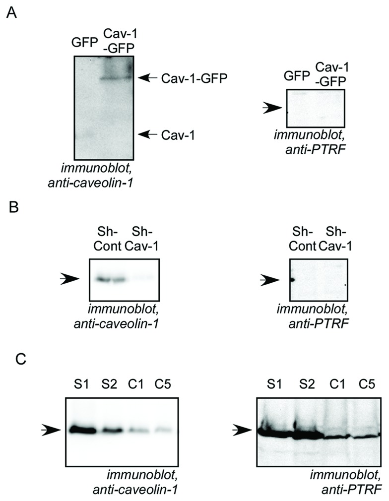
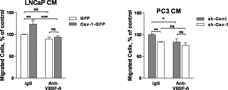
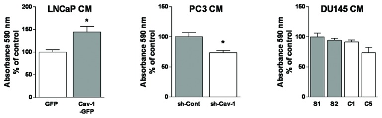
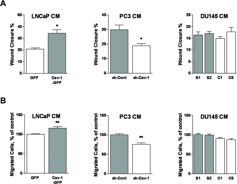
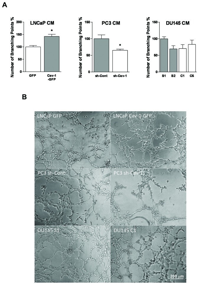
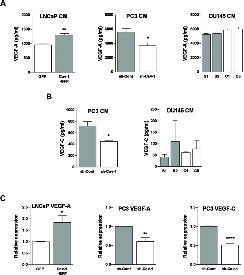
Lymphangiogenesis allows prostate cancer (PCa) lymphatic metastasis, which is associated with poor prognosis and short survival rates. Caveolin-1 (Cav-1) is a membrane protein localized in caveolae, but also exists in non-caveolar, cellular or extracellular forms. Cav-1 is overexpressed in PCa, promotes prostate tumour progression and metastasis. We investigated the effect of caveolar and non-caveolar Cav-1 on PCa lymphangiogenic potential. Cav-1 was down-regulated in PC3 and DU145, and ectopically expressed in LNCaP cells. The effect of PCa cell conditioned media on lymphatic endothelial cell (LEC) viability, chemotaxis, chemokinesis and differentiation was assessed. The effect of Cav-1 on PCa cell expression of lymphangiogenesis-modulators VEGF-A and VEGF-C was assessed using qPCR and ELISA of the conditioned medium. Non-caveolar Cav-1, whether exogenous or endogenous (in LNCaP and PC3 cells, respectively) enhanced LEC proliferation, migration and differentiation. In contrast, caveolar Cav-1 (in DU145 cells) did not significantly affect PCa cell lymphangiogenic potential. The effect of non-caveolar Cav-1 on LECs was mediated by increased expression of VEGF-A as demonstrated by neutralization by anti-VEGF-A antibody. This study unveils for the first time a crucial role for non-caveolar Cav-1 in modulating PCa cell expression of VEGF-A and subsequent LEC proliferation, migration and tube formation.
淋巴管生成促使前列腺癌发生淋巴转移,这与预后不良和生存率低相关。小窝蛋白-1(Cav-1)是一种定位于小窝的膜蛋白,但也以非小窝、细胞内或细胞外形式存在。Cav-1在前列腺癌中过度表达,促进前列腺肿瘤进展和转移。我们研究了小窝型和非小窝型Cav-1对前列腺癌细胞淋巴管生成潜能的影响。在PC3和DU145细胞中下调Cav-1,并在LNCaP细胞中异位表达。评估前列腺癌细胞条件培养基对淋巴管内皮细胞(LEC)活力、趋化性、化学运动性和分化的影响。使用条件培养基的qPCR和ELISA评估Cav-1对前列腺癌细胞淋巴管生成调节因子VEGF-A和VEGF-C表达的影响。非小窝型Cav-1,无论是外源性的还是内源性的(分别在LNCaP和PC3细胞中),均可增强LEC的增殖、迁移和分化。相比之下,小窝型Cav-1(在DU145细胞中)对前列腺癌细胞淋巴管生成潜能没有显著影响。抗VEGF-A抗体中和实验表明,非小窝型Cav-1对LEC的作用是通过增加VEGF-A的表达介导的。本研究首次揭示了非小窝型Cav-1在调节前列腺癌细胞VEGF-A表达以及随后的LEC增殖、迁移和管腔形成中的关键作用。