Disdier Clémence, Devoy Jérôme, Cosnefroy Anne, Chalansonnet Monique, Herlin-Boime Nathalie, Brun Emilie, Lund Amie, Mabondzo Aloïse
CEA, Direction des Sciences du Vivant, iBiTec-S, Service de Pharmacologie et d'Immunoanalyse, Equipe Pharmacologie Neurovasculaire, 91191, Gif-sur-Yvette, France.
INRS, Département Polluants et Santé, Rue du Morvan, CS 60027, 54519, Vandœuvre Cedex, France.
Part Fibre Toxicol. 2015 Sep 4;12:27. doi: 10.1186/s12989-015-0102-8.
Notwithstanding increasing knowledge of titanium dioxide nanoparticles (TiO2 NPs) passing through biological barriers, their biodistribution to the central nervous system (CNS) and potential effects on blood-brain barrier (BBB) physiology remain poorly characterized.
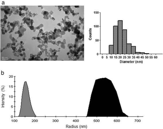
Here, we report time-related responses from single-dose intravenous (IV) administration of 1 mg/kg TiO2 NPs to rats, with particular emphasis on titanium (Ti) quantification in the brain. Ti content in tissues was analyzed using inductively coupled plasma mass spectrometry. Integrity and functionality of the BBB as well as brain inflammation were characterized using a panel of methods including RT-PCR, immuno-histo chemistry and transporter activity evaluation.
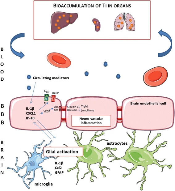
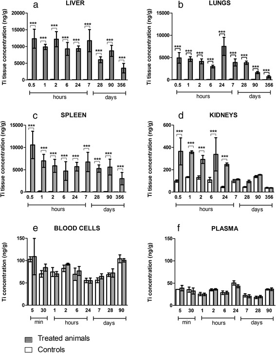
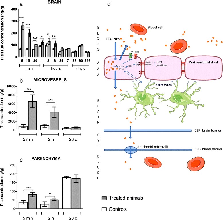
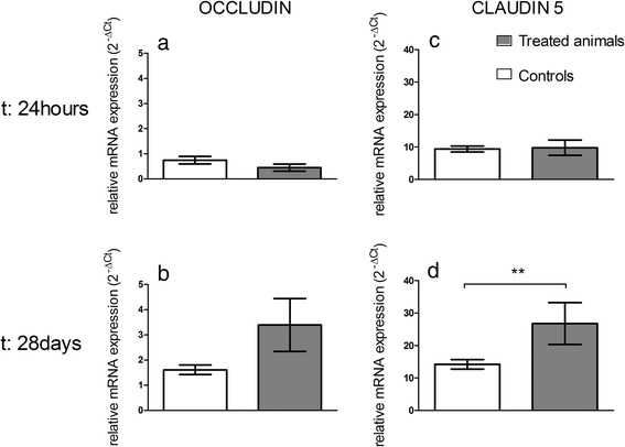
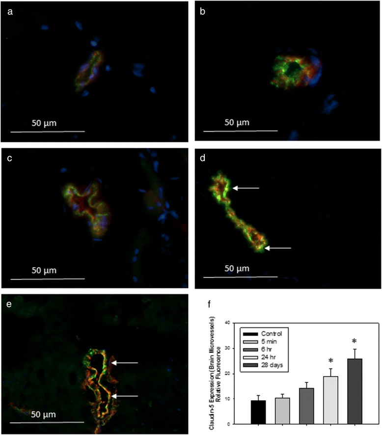
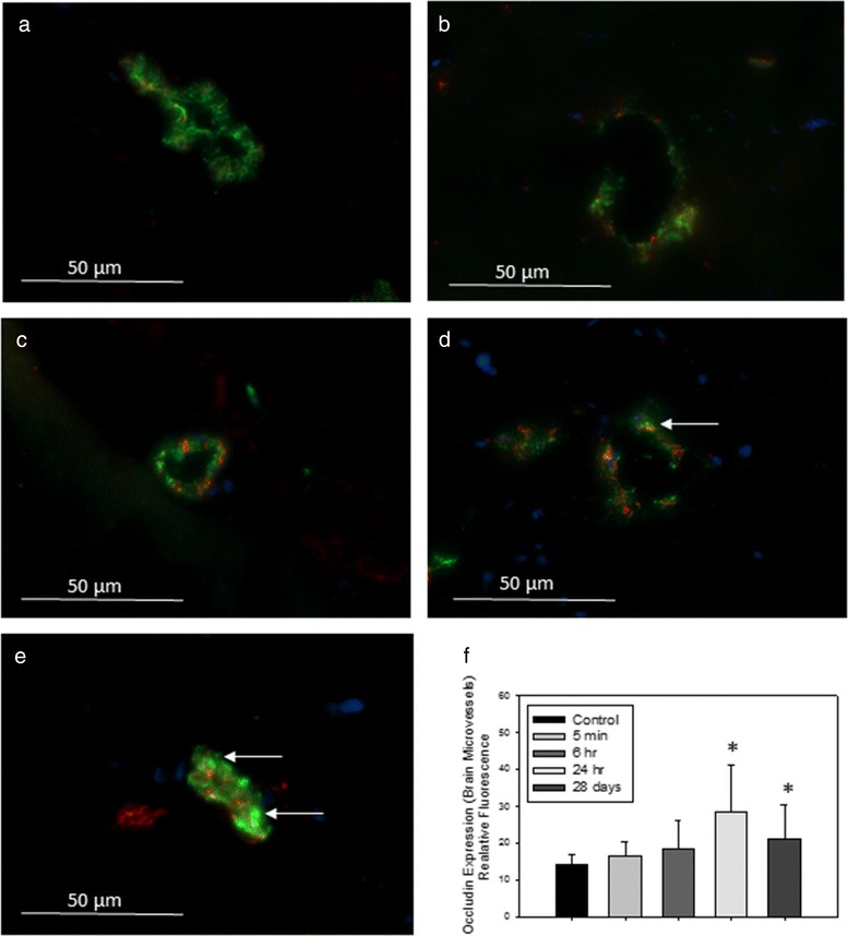
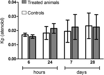
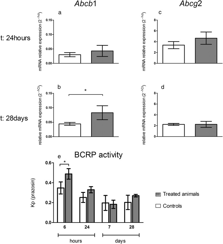
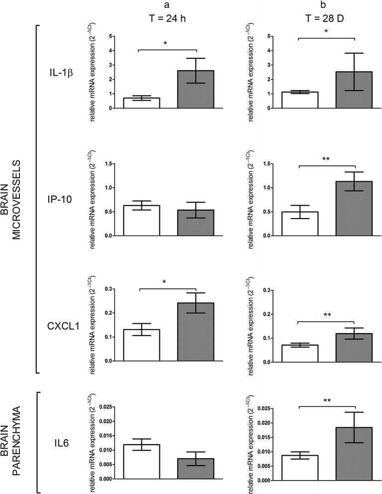
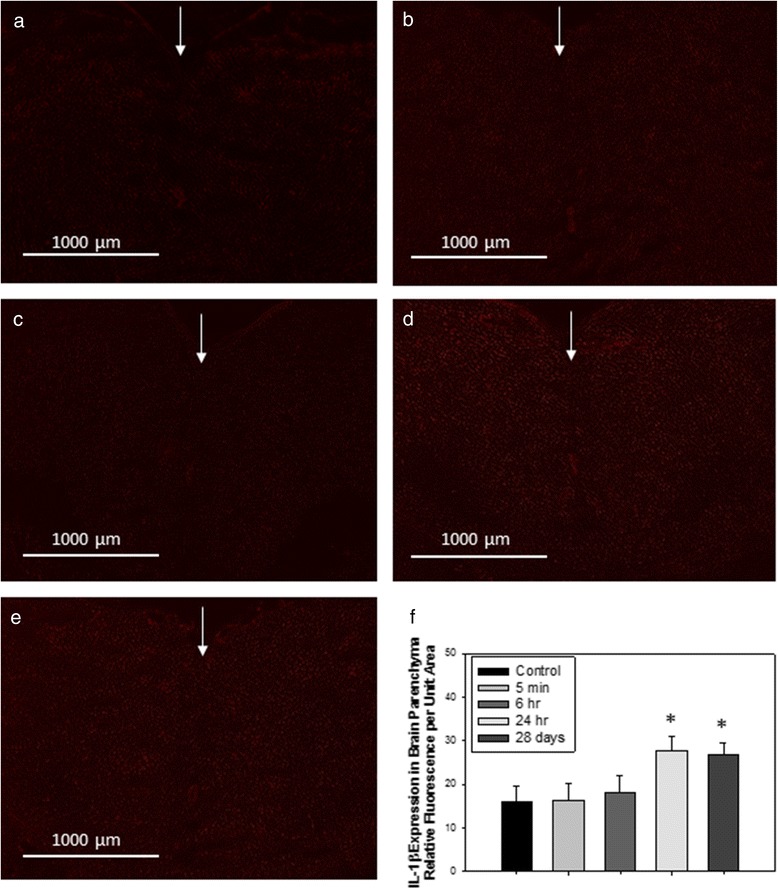
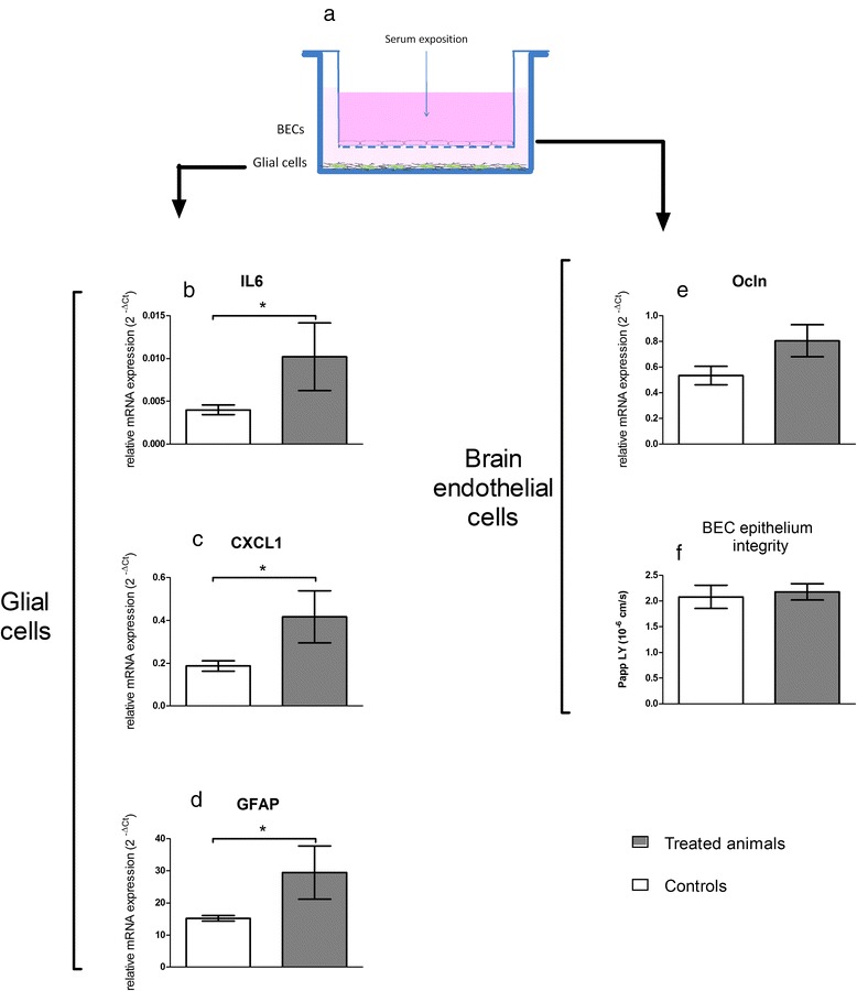
Biokinetic analysis revealed Ti biopersistence in liver, lungs and spleen up to one year after TiO2 NPs administration. A significant increase of Ti in the brain was observed at early end points followed by a subsequent decrease. In-depth analysis of Ti in the total brain demonstrated quantitative Ti uptake and clearance by brain microvasculature endothelial cells (BECs) with minimal translocation in the brain parenchyma. The presence of Ti in the BECs did not affect BBB integrity, despite rapid reversible modulation of breast cancer resistance protein activity. Ti biopersistence in organs such as liver was associated with significant increases of tight junction proteins (claudin-5 and occludin), interleukin 1β (IL-1β), chemokine ligand 1 (CXCL1) and γ inducible protein-10 (IP-10/CXCL10) in BECs and also increased levels of IL-1β in brain parenchyma despite lack of evidence of Ti in the brain. These findings mentioned suggest potential effect of Ti present at a distance from the brain possibly via mediators transported by blood. Exposure of an in vitro BBB model to sera from TiO2 NPs-treated animals confirmed the tightness of the BBB and inflammatory responses.
Overall, these findings suggest the clearance of TiO2 NPs at the BBB with persistent brain inflammation and underscore the role of Ti biopersistence in organs that can exert indirect effects on the CNS dependent on circulating factors.
尽管对二氧化钛纳米颗粒(TiO₂ NPs)穿过生物屏障的了解日益增加,但其在中枢神经系统(CNS)中的生物分布以及对血脑屏障(BBB)生理学的潜在影响仍知之甚少。
在此,我们报告了对大鼠单次静脉注射(IV)1 mg/kg TiO₂ NPs后的时间相关反应,特别强调了脑中钛(Ti)的定量分析。使用电感耦合等离子体质谱法分析组织中的Ti含量。使用包括RT-PCR、免疫组织化学和转运体活性评估在内的一系列方法来表征BBB的完整性和功能以及脑内炎症。
生物动力学分析显示,TiO₂ NPs给药后长达一年,Ti在肝脏、肺和脾脏中具有生物持久性。在早期时间点观察到脑中Ti显著增加,随后减少。对全脑中Ti的深入分析表明,脑微血管内皮细胞(BECs)对Ti有定量摄取和清除,而在脑实质中的转运极少。尽管乳腺癌耐药蛋白活性有快速可逆的调节,但BECs中Ti的存在并未影响BBB的完整性。肝脏等器官中Ti的生物持久性与BECs中紧密连接蛋白(claudin-5和occludin)、白细胞介素1β(IL-1β)、趋化因子配体1(CXCL1)和γ诱导蛋白10(IP-10/CXCL10)的显著增加有关,尽管脑中没有Ti的证据,但脑实质中IL-1β水平也有所升高。上述发现表明,远离大脑的Ti可能通过血液运输的介质产生潜在影响。将体外BBB模型暴露于TiO₂ NPs处理动物的血清中证实了BBB的紧密性和炎症反应。
总体而言,这些发现表明TiO₂ NPs在BBB处被清除,同时伴有持续性脑内炎症,并强调了Ti在器官中的生物持久性作用,这种作用可能通过循环因子对中枢神经系统产生间接影响。