Choi Soo-Young, Jeong Woo-Chang, Lee Young-Won, Choi Ho-Jung
College of Veterinary Medicine and Research Institute of Veterinary Medicine, Chungnam National University, Daejeon 305-764, South Korea.
J Vet Med Sci. 2016 Feb;78(2):239-44. doi: 10.1292/jvms.15-0199. Epub 2015 Sep 28.
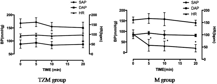
Contrast enhanced ultrasound (CEUS) is useful to evaluate tissue perfusion in the kidney. In veterinary medicine, sedation or anesthesia may be required in uncooperative or panting patients. The aim of this study was to evaluate and compare the normal kidney perfusion patterns in conscious and anesthetized dogs using CEUS. Eight healthy beagles were used in this study. Scanning was performed in conscious dogs using manual restraint (conscious group), or under general anesthesia using tiletamine-zolazepam and medetomidine (TZM group) or medetomidine (M group). The contrast agent (Sonovue(®)) was administered as an IV bolus. The peak intensity (PI), time to peak enhancement from injection (TTP0) and the time to peak enhancement from the initial rise (TTPup), upslope, downslope and area under the curve (AUC) were analyzed. Compared to the cortical values in the conscious group, TTP0 was significantly delayed in the TZM group, and upslope, TTP0 and TTPup were significantly different in the M group. The AUCs in the TZM and M groups were not different from those in the conscious group. The upslope of renal medullary perfusion was significantly decreased in the TZM and M groups. TTP0 and TTPup were also significantly delayed in these groups. The AUC of the medulla was significantly decreased in the M group. Therefore, TZM is useful as an anesthetic protocol when performing CEUS, and the obtained data may serve as reference values in the evaluation of renal perfusion using CEUS in dogs under anesthesia.
超声造影(CEUS)有助于评估肾脏的组织灌注。在兽医学中,对于不配合或喘气的患者可能需要镇静或麻醉。本研究的目的是使用CEUS评估和比较清醒和麻醉犬的正常肾脏灌注模式。本研究使用了8只健康的比格犬。在清醒犬中使用手动约束进行扫描(清醒组),或在使用替来他明-唑拉西泮和美托咪定(TZM组)或美托咪定(M组)进行全身麻醉下扫描。造影剂(声诺维(®))作为静脉推注给药。分析了峰值强度(PI)、注射后达到峰值增强的时间(TTP0)、从初始上升到峰值增强的时间(TTPup)、上升斜率、下降斜率和曲线下面积(AUC)。与清醒组的皮质值相比,TZM组的TTP0显著延迟,M组的上升斜率、TTP0和TTPup显著不同。TZM组和M组的AUC与清醒组无差异。TZM组和M组肾髓质灌注的上升斜率显著降低。这些组的TTP0和TTPup也显著延迟。M组髓质的AUC显著降低。因此,TZM在进行CEUS时作为一种麻醉方案是有用的,并且所获得的数据可作为麻醉下犬使用CEUS评估肾脏灌注的参考值。