Stoehr Linda C, Endes Carola, Radauer-Preiml Isabella, Boyles Matthew S P, Casals Eudald, Balog Sandor, Pesch Markus, Petri-Fink Alke, Rothen-Rutishauser Barbara, Himly Martin, Clift Martin J D, Duschl Albert
Department of Molecular Biology, University of Salzburg, Hellbrunnerstrasse 34, 5020, Salzburg, Austria.
Grimm Aerosol Technik GmbH & Co. KG, Ainring, Germany.
Part Fibre Toxicol. 2015 Sep 29;12:29. doi: 10.1186/s12989-015-0104-6.
Stably transfected lung epithelial reporter cell lines pose an advantageous alternative to replace complex experimental techniques to monitor the pro-inflammatory response following nanoparticle (NP) exposure. Previously, reporter cell lines have been used under submerged culture conditions, however, their potential usefulness in combination with air-liquid interface (ALI) exposures is currently unknown. Therefore, the aim of the present study was to compare a panel of interleukin-8 promoter (pIL8)-reporter cell lines (i.e. green or red fluorescent protein (GFP, RFP), and luciferase (Luc)), originating from A549 lung epithelial type II-like cells cells, following NPs exposure under both submerged and ALI conditions.
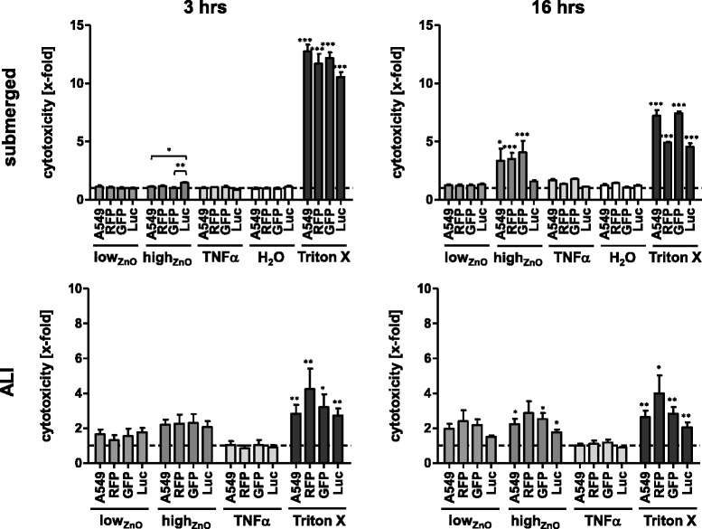
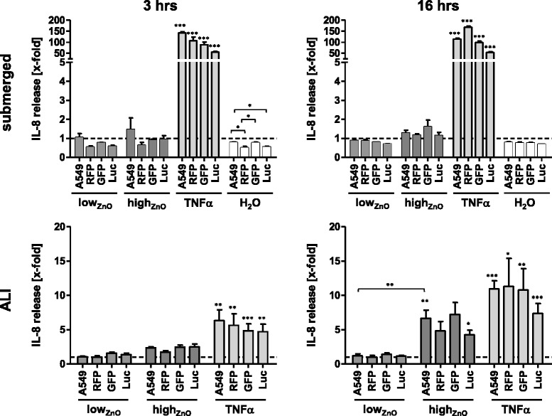
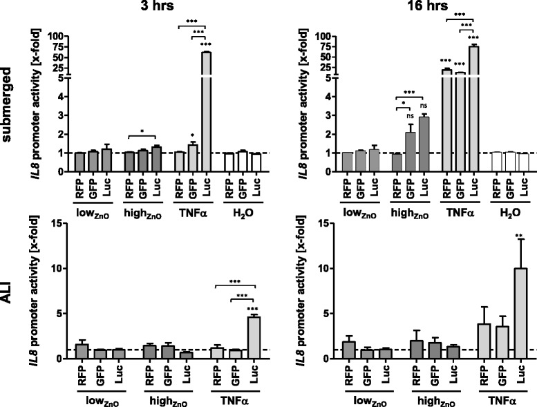
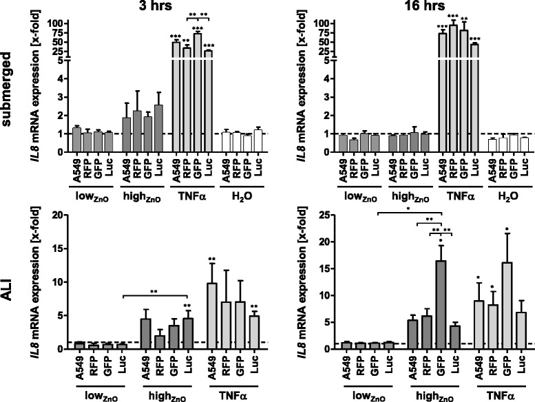
All cell lines were exposed to zinc oxide (ZnO) NPs at 0.6 and 6.2 μg/cm(2) for 3 and 16 hours under both submerged and ALI conditions. Following physicochemical characterization, the cytotoxic profile of the ZnO-NPs was determined for each exposure scenario. Expression of IL-8 from all cell types was analyzed at the promoter level and compared to the mRNA (qRT-PCR) and protein level (ELISA).
In summary, each reporter cell line detected acute pro-inflammatory effects following ZnO exposure under each condition tested. The pIL8-Luc cell line was the most sensitive in terms of reporter signal strength and onset velocity following TNF-α treatment. Both pIL8-GFP and pIL8-RFP also showed a marked signal induction in response to TNF-α, although only after 16 hrs. In terms of ZnO-NP-induced cytotoxicity pIL8-RFP cells were the most affected, whilst the pIL8-Luc were found the least responsive.
In conclusion, the use of fluorescence-based reporter cell lines can provide a useful tool in screening the pro-inflammatory response following NP exposure in both submerged and ALI cell cultures.
稳定转染的肺上皮报告细胞系是一种有利的替代方法,可取代复杂的实验技术来监测纳米颗粒(NP)暴露后的促炎反应。此前,报告细胞系已用于浸没培养条件下,然而,其与气液界面(ALI)暴露联合使用的潜在效用目前尚不清楚。因此,本研究的目的是比较一组源自A549 II型肺上皮样细胞的白细胞介素-8启动子(pIL8)-报告细胞系(即绿色或红色荧光蛋白(GFP、RFP)和荧光素酶(Luc))在浸没和ALI条件下NP暴露后的情况。
所有细胞系在浸没和ALI条件下,分别以0.6和6.2μg/cm²的剂量暴露于氧化锌(ZnO)纳米颗粒3小时和16小时。在进行物理化学表征后,确定每种暴露情况下ZnO纳米颗粒的细胞毒性特征。分析所有细胞类型中IL-8在启动子水平的表达,并与mRNA(qRT-PCR)和蛋白质水平(ELISA)进行比较。
总之,在每种测试条件下,每种报告细胞系均检测到ZnO暴露后的急性促炎作用。就报告信号强度和TNF-α处理后的起始速度而言,pIL8-Luc细胞系最为敏感。pIL8-GFP和pIL8-RFP在TNF-α刺激下也显示出明显的信号诱导,不过仅在16小时后。就ZnO纳米颗粒诱导的细胞毒性而言,pIL8-RFP细胞受影响最大,而pIL8-Luc细胞反应最小。
总之,基于荧光的报告细胞系的使用可为筛选浸没和ALI细胞培养中NP暴露后的促炎反应提供有用工具。