Wang Hui, Li Bei, Feng Yanmei, Cui Biao, Wu Hongmin, Shi Haibo, Yin Shankai
Department of Otolaryngology head and neck surgery, Shanghai Jiao Tong University Affiliated Sixth People's Hospital, Shanghai, China, 200233.
PLoS One. 2015 Oct 2;10(10):e0139622. doi: 10.1371/journal.pone.0139622. eCollection 2015.
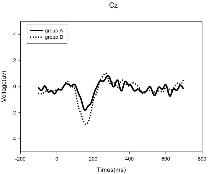
Repetitive Transcranial Magnetic Stimulation (rTMS) is a novel therapeutic tool to induce a suppression of tinnitus. However, the optimal target sites are unknown. We aimed to determine whether low-frequency rTMS induced lasting suppression of tinnitus by decreasing neural activity in the cortex, navigated by high-density electroencephalogram (EEG) source analysis, and the utility of EEG for targeting treatment.
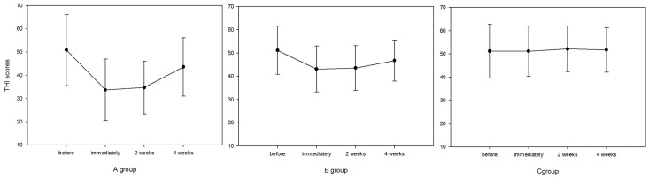
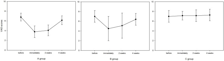
In this controlled three-armed trial, seven normal hearing patients with tonal tinnitus received a 10-day course of 1-Hz rTMS to the cortex, navigated by high-density EEG source analysis, to the left temporoparietal cortex region, and to the left temporoparietal with sham stimulation. The Tinnitus handicap inventory (THI) and a visual analog scale (VAS) were used to assess tinnitus severity and loudness. Measurements were taken before, and immediately, 2 weeks, and 4 weeks after the end of the interventions.
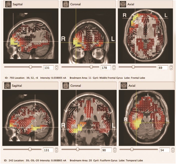
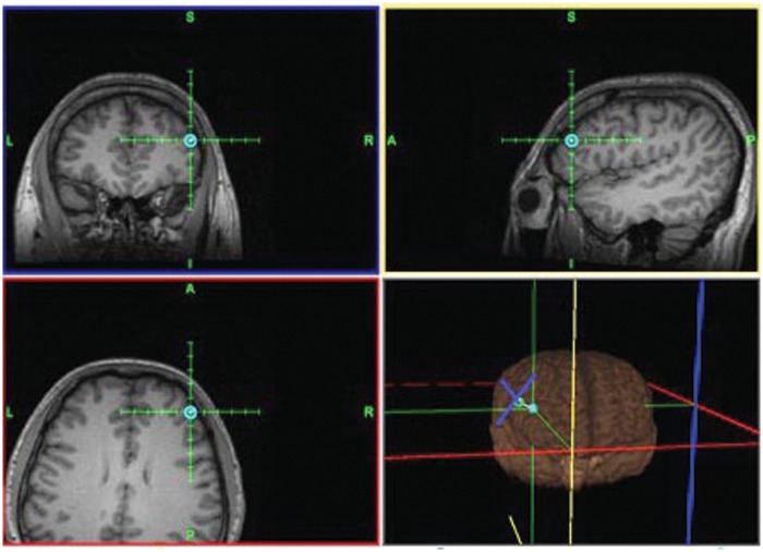
Low-frequency rTMS decreased tinnitus significantly after active, but not sham, treatment. Responders in the EEG source analysis-based rTMS group, 71.4% (5/7) patients, experienced a significant reduction in tinnitus loudness, as evidenced by VAS scores. The target site of neuronal generators most consistently associated with a positive response was the frontal lobe in the right hemisphere, sourced using high-density EEG equipment, in the tinnitus patients. After left temporoparietal rTMS stimulation, 42.8% (3/7) patients experienced a decrease in tinnitus loudness.
Active EEG source analysis based rTMS resulted in significant suppression in tinnitus loudness, showing the superiority of neuronavigation-guided coil positioning in dealing with tinnitus. Non-auditory areas should be considered in the pathophysiology of tinnitus. This knowledge in turn can contribute to investigate the pathophysiology of tinnitus.
重复经颅磁刺激(rTMS)是一种诱导耳鸣抑制的新型治疗工具。然而,最佳靶点位置尚不清楚。我们旨在通过高密度脑电图(EEG)源分析引导,确定低频rTMS是否通过降低皮层神经活动诱导耳鸣的持续抑制,以及EEG在靶向治疗中的效用。
在这项对照三臂试验中,7名患有音调性耳鸣的听力正常患者接受了为期10天的1赫兹rTMS治疗,通过高密度EEG源分析引导,刺激左侧颞顶叶皮层区域,以及对左侧颞顶叶进行假刺激。使用耳鸣障碍量表(THI)和视觉模拟量表(VAS)评估耳鸣严重程度和响度。在干预结束前、结束后即刻、2周和4周进行测量。
主动治疗而非假治疗后,低频rTMS显著降低了耳鸣。基于EEG源分析的rTMS组中的反应者,即71.4%(5/7)的患者,耳鸣响度显著降低,VAS评分证明了这一点。在耳鸣患者中,使用高密度EEG设备确定,与阳性反应最一致相关的神经元发生器靶点位于右半球额叶。左侧颞顶叶rTMS刺激后,42.8%(3/7)的患者耳鸣响度降低。
基于主动EEG源分析的rTMS导致耳鸣响度显著抑制,显示了神经导航引导线圈定位在治疗耳鸣方面的优越性。耳鸣的病理生理学应考虑非听觉区域。这一知识反过来有助于研究耳鸣的病理生理学。