Piotrowski Wojciech J, Kiszałkiewicz Justyna, Górski Paweł, Antczak Adam, Górski Witold, Pastuszak-Lewandoska Dorota, Migdalska-Sęk Monika, Domańska-Senderowska Daria, Nawrot Ewa, Czarnecka Karolina H, Kurmanowska Zofia, Brzeziańska-Lasota Ewa
Department of Pneumonology and Allergy, 1st Chair of Internal Medicine, Medical University of Lodz, Lodz, Poland.
Department of Molecular Bases of Medicine, 1st Chair of Internal Medicine, Medical University of Lodz, 251 Pomorska St., 92-213, Lodz, Poland.
BMC Immunol. 2015 Oct 6;16:58. doi: 10.1186/s12865-015-0123-y.
The chronic course of pulmonary sarcoidosis can lead to lung dysfunction due to fibrosis, in which the signalling pathways TGF-β/Smad and VEGF-A may play a key role.
We evaluated immunoexpression of TGF-β1, Smad2, 3, and 7, and VEGF-A in serum and bronchoalveolar lavage (BAL) fluid of patients (n = 57) classified according to the presence of lung parenchymal involvement (radiological stage I vs. II-III), acute vs. insidious onset, lung function test (LFT) results, calcium metabolism parameters, percentage of BAL lymphocytes (BAL-L%), BAL CD4(+)/CD8(+) ratio, age, and gender. Immunoexpression analysis of proteins was performed by ELISA.
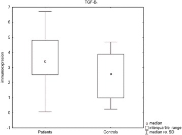
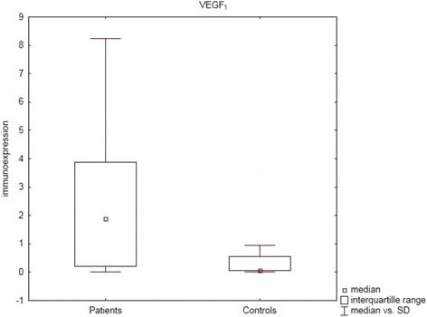
The immunoexpression of all studied proteins were higher in serum than in BAL fluid of patients (p >0.05). The serum levels of TGF-β1 (p = 0.03), Smad2 (p = 0.01), and VEGF-A (p = 0.0002) were significantly higher in sarcoidosis patients compared to healthy controls. There were no differences within the sarcoidosis group between patients with vs. without parenchymal involvement, acute vs. insidious onset, or patients with normal vs. abnormal spirometry results. In patients with abnormal spirometry results a negative correlation was found between forced vital capacity (FVC) % predicted value and TGF-β1 immunoexpression in BAL fluid, and positive correlations were observed between the intensity of lung parenchymal changes estimated by high-resolution computed tomography (HRCT scores) and Smad 2 level in serum.
TGF-β/Smad signalling pathway and VEGF-A participate in the pathogenesis of sarcoidosis. BAL TGF-β1, and Smad 2 in serum seem to be promising biomarkers with negative prognostic value, but further studies are required to confirmed our observations.
肺结节病的慢性病程可因纤维化导致肺功能障碍,其中转化生长因子-β(TGF-β)/Smad信号通路和血管内皮生长因子-A(VEGF-A)可能起关键作用。
我们评估了57例患者血清和支气管肺泡灌洗(BAL)液中TGF-β1、Smad2、3和7以及VEGF-A的免疫表达,这些患者根据肺实质受累情况(放射学分期I期与II - III期)、急性与隐匿性起病、肺功能测试(LFT)结果、钙代谢参数、BAL淋巴细胞百分比(BAL-L%)、BAL CD4(+)/CD8(+)比值、年龄和性别进行分类。通过酶联免疫吸附测定(ELISA)进行蛋白质的免疫表达分析。
所有研究蛋白的免疫表达在患者血清中均高于BAL液(p>0.05)。与健康对照相比,结节病患者血清中TGF-β1(p = 0.03)、Smad2(p = 0.01)和VEGF-A(p = 0.0002)水平显著更高。在结节病组中,有或无实质受累的患者、急性与隐匿性起病的患者或肺量计结果正常与异常的患者之间无差异。在肺量计结果异常的患者中,发现BAL液中用力肺活量(FVC)预测值百分比与TGF-β1免疫表达呈负相关,血清中Smad 2水平与高分辨率计算机断层扫描(HRCT评分)估计的肺实质改变强度呈正相关。
TGF-β/Smad信号通路和VEGF-A参与结节病的发病机制。BAL中的TGF-β1和血清中的Smad 2似乎是具有负面预后价值的有前景的生物标志物,但需要进一步研究来证实我们的观察结果。