• 文献检索
  • 文档翻译
  • 深度研究
  • 学术资讯
  • Suppr Zotero 插件Zotero 插件
  • 邀请有礼
  • 套餐&价格
  • 历史记录
应用&插件
Suppr Zotero 插件Zotero 插件浏览器插件Mac 客户端Windows 客户端微信小程序
定价
高级版会员购买积分包购买API积分包
服务
文献检索文档翻译深度研究API 文档MCP 服务
关于我们
关于 Suppr公司介绍联系我们用户协议隐私条款
关注我们

Suppr 超能文献

核心技术专利:CN118964589B侵权必究
粤ICP备2023148730 号-1Suppr @ 2026

文献检索

告别复杂PubMed语法,用中文像聊天一样搜索,搜遍4000万医学文献。AI智能推荐,让科研检索更轻松。

立即免费搜索

文件翻译

保留排版,准确专业,支持PDF/Word/PPT等文件格式,支持 12+语言互译。

免费翻译文档

深度研究

AI帮你快速写综述,25分钟生成高质量综述,智能提取关键信息,辅助科研写作。

立即免费体验

基于滑动滤波器的神经元解剖结构重建

Neuron anatomy structure reconstruction based on a sliding filter.

作者信息

Luo Gongning, Sui Dong, Wang Kuanquan, Chae Jinseok

机构信息

Research Center of Perception and Computing, School of Computer Science and Technology, Harbin Institute of Technology, Harbin, China.

Department of Computer Science and Engineering, Incheon National University, Incheon, Korea.

出版信息

BMC Bioinformatics. 2015 Oct 24;16:342. doi: 10.1186/s12859-015-0780-0.

DOI:10.1186/s12859-015-0780-0
PMID:26498293
原文链接:https://pmc.ncbi.nlm.nih.gov/articles/PMC4619512/
Abstract

BACKGROUND

Reconstruction of neuron anatomy structure is a challenging and important task in neuroscience. However, few algorithms can automatically reconstruct the full structure well without manual assistance, making it essential to develop new methods for this task.

METHODS

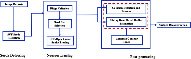
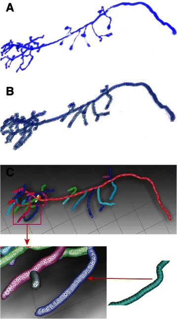
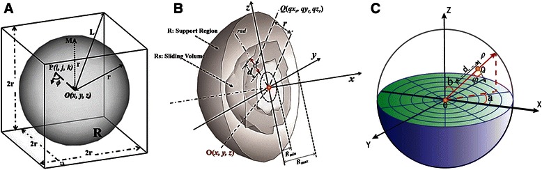
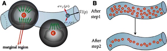
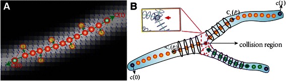
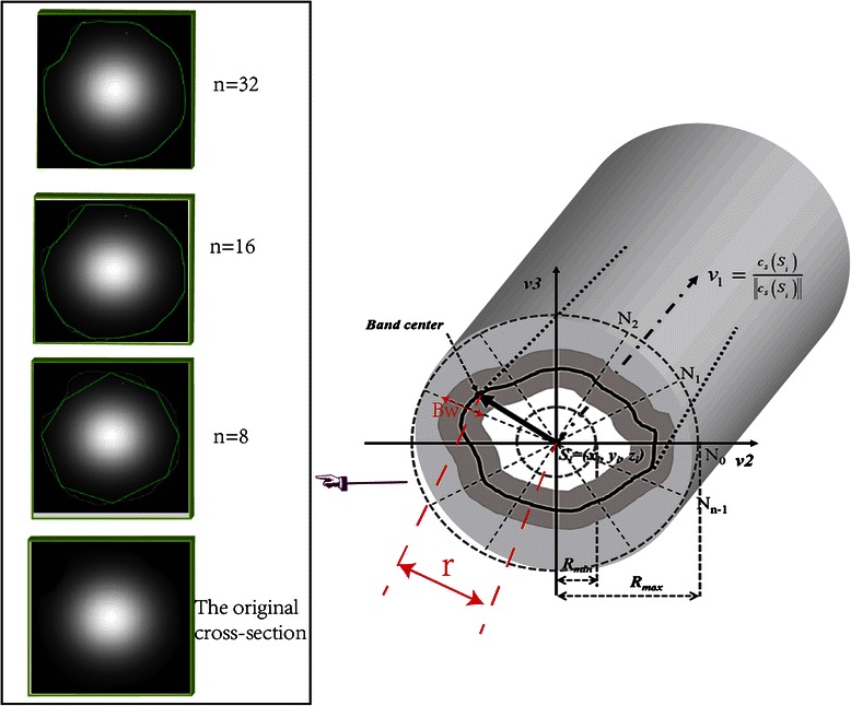
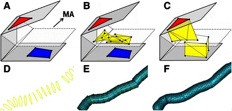
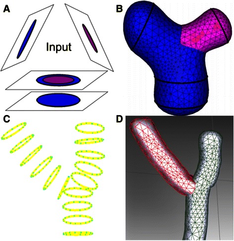
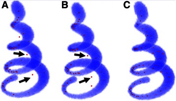
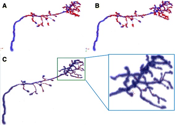
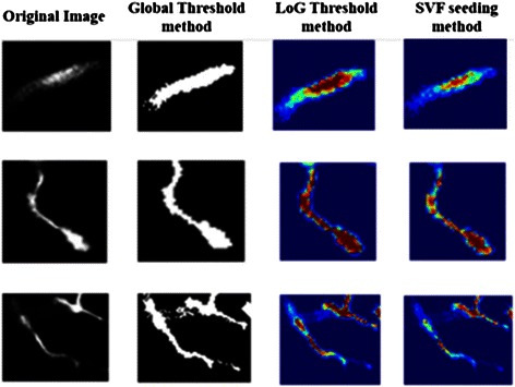
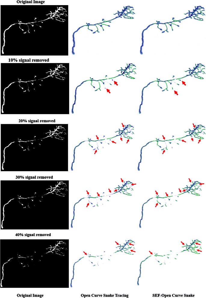
This paper introduces a new pipeline for reconstructing neuron anatomy structure from 3-D microscopy image stacks. This pipeline is initialized with a set of seeds that were detected by our proposed Sliding Volume Filter (SVF), given a non-circular cross-section of a neuron cell. Then, an improved open curve snake model combined with a SVF external force is applied to trace the full skeleton of the neuron cell. A radius estimation method based on a 2D sliding band filter is developed to fit the real edge of the cross-section of the neuron cell. Finally, a surface reconstruction method based on non-parallel curve networks is used to generate the neuron cell surface to finish this pipeline.

RESULTS

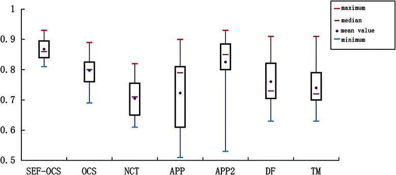
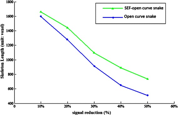
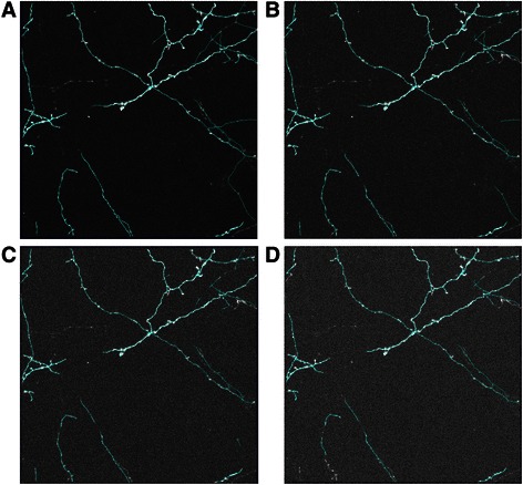
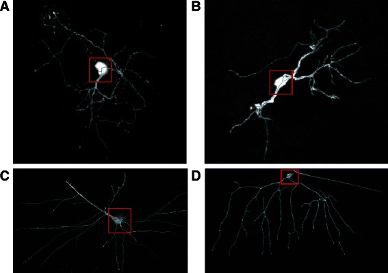
The proposed pipeline has been evaluated using publicly available datasets. The results show that the proposed method achieves promising results in some datasets from the DIgital reconstruction of Axonal and DEndritic Morphology (DIADEM) challenge and new BigNeuron project.

CONCLUSION

The new pipeline works well in neuron tracing and reconstruction. It can achieve higher efficiency, stability and robustness in neuron skeleton tracing. Furthermore, the proposed radius estimation method and applied surface reconstruction method can obtain more accurate neuron anatomy structures.

摘要

背景

神经元解剖结构的重建是神经科学中一项具有挑战性且重要的任务。然而,几乎没有算法能够在没有人工辅助的情况下自动很好地重建完整结构,因此开发针对此任务的新方法至关重要。

方法

本文介绍了一种从三维显微镜图像堆栈重建神经元解剖结构的新流程。给定神经元细胞的非圆形横截面,该流程以通过我们提出的滑动体积滤波器(SVF)检测到的一组种子点进行初始化。然后,应用一种改进的开放曲线蛇模型并结合SVF外力来追踪神经元细胞的完整骨架。开发了一种基于二维滑动带滤波器的半径估计方法,以拟合神经元细胞横截面的真实边缘。最后,使用基于非平行曲线网络的表面重建方法生成神经元细胞表面,从而完成该流程。

结果

已使用公开可用的数据集对所提出的流程进行了评估。结果表明,在轴突和树突形态的数字重建(DIADEM)挑战和新的大神经元项目的一些数据集中,所提出的方法取得了有前景的结果。

结论

新流程在神经元追踪和重建方面效果良好。它在神经元骨架追踪中可以实现更高的效率、稳定性和鲁棒性。此外,所提出的半径估计方法和应用的表面重建方法能够获得更准确的神经元解剖结构。

https://cdn.ncbi.nlm.nih.gov/pmc/blobs/bd84/4619512/13f5372c07f2/12859_2015_780_Fig17_HTML.jpg
https://cdn.ncbi.nlm.nih.gov/pmc/blobs/bd84/4619512/3ffccd5d4ddf/12859_2015_780_Fig1_HTML.jpg
https://cdn.ncbi.nlm.nih.gov/pmc/blobs/bd84/4619512/a2b126c2efd0/12859_2015_780_Fig2_HTML.jpg
https://cdn.ncbi.nlm.nih.gov/pmc/blobs/bd84/4619512/65466b9300ae/12859_2015_780_Fig3_HTML.jpg
https://cdn.ncbi.nlm.nih.gov/pmc/blobs/bd84/4619512/32bf22ff67d9/12859_2015_780_Fig4_HTML.jpg
https://cdn.ncbi.nlm.nih.gov/pmc/blobs/bd84/4619512/b2d8bd48fbd4/12859_2015_780_Fig5_HTML.jpg
https://cdn.ncbi.nlm.nih.gov/pmc/blobs/bd84/4619512/853cc81d14ab/12859_2015_780_Fig6_HTML.jpg
https://cdn.ncbi.nlm.nih.gov/pmc/blobs/bd84/4619512/f9d247e01559/12859_2015_780_Fig7_HTML.jpg
https://cdn.ncbi.nlm.nih.gov/pmc/blobs/bd84/4619512/5c3babde23f2/12859_2015_780_Fig8_HTML.jpg
https://cdn.ncbi.nlm.nih.gov/pmc/blobs/bd84/4619512/fb765b7ea77f/12859_2015_780_Fig9_HTML.jpg
https://cdn.ncbi.nlm.nih.gov/pmc/blobs/bd84/4619512/1f3dadc6fce8/12859_2015_780_Fig10_HTML.jpg
https://cdn.ncbi.nlm.nih.gov/pmc/blobs/bd84/4619512/207a9db53f33/12859_2015_780_Fig11_HTML.jpg
https://cdn.ncbi.nlm.nih.gov/pmc/blobs/bd84/4619512/8ff94804320a/12859_2015_780_Fig12_HTML.jpg
https://cdn.ncbi.nlm.nih.gov/pmc/blobs/bd84/4619512/7c80e199f958/12859_2015_780_Fig13_HTML.jpg
https://cdn.ncbi.nlm.nih.gov/pmc/blobs/bd84/4619512/6962f2809f08/12859_2015_780_Fig14_HTML.jpg
https://cdn.ncbi.nlm.nih.gov/pmc/blobs/bd84/4619512/be692cac2432/12859_2015_780_Fig15_HTML.jpg
https://cdn.ncbi.nlm.nih.gov/pmc/blobs/bd84/4619512/528dc64adede/12859_2015_780_Fig16_HTML.jpg
https://cdn.ncbi.nlm.nih.gov/pmc/blobs/bd84/4619512/13f5372c07f2/12859_2015_780_Fig17_HTML.jpg

相似文献

1
Neuron anatomy structure reconstruction based on a sliding filter.基于滑动滤波器的神经元解剖结构重建
BMC Bioinformatics. 2015 Oct 24;16:342. doi: 10.1186/s12859-015-0780-0.
2
A pipeline for neuron reconstruction based on spatial sliding volume filter seeding.一种基于空间滑动体积滤波器种子点的神经元重建流水线。
Comput Math Methods Med. 2014;2014:386974. doi: 10.1155/2014/386974. Epub 2014 Jul 2.
3
Rivulet: 3D Neuron Morphology Tracing with Iterative Back-Tracking.Rivulet:基于迭代回溯的3D神经元形态追踪
Neuroinformatics. 2016 Oct;14(4):387-401. doi: 10.1007/s12021-016-9302-0.
4
SparseTracer: the Reconstruction of Discontinuous Neuronal Morphology in Noisy Images.稀疏追踪器:噪声图像中不连续神经元形态的重建
Neuroinformatics. 2017 Apr;15(2):133-149. doi: 10.1007/s12021-016-9317-6.
5
NRTR: Neuron Reconstruction With Transformer From 3D Optical Microscopy Images.NRTR:基于 3D 光学显微镜图像的 Transformer 神经元重建。
IEEE Trans Med Imaging. 2024 Feb;43(2):886-898. doi: 10.1109/TMI.2023.3323466. Epub 2024 Feb 2.
6
BigNeuron: a resource to benchmark and predict performance of algorithms for automated tracing of neurons in light microscopy datasets.BigNeuron:一个基准资源,用于评估和预测在光显微镜数据集上自动追踪神经元的算法的性能。
Nat Methods. 2023 Jun;20(6):824-835. doi: 10.1038/s41592-023-01848-5. Epub 2023 Apr 17.
7
A broadly applicable 3-D neuron tracing method based on open-curve snake.基于开曲线蛇的一种广泛适用的三维神经元示踪方法。
Neuroinformatics. 2011 Sep;9(2-3):193-217. doi: 10.1007/s12021-011-9110-5.
8
M-AMST: an automatic 3D neuron tracing method based on mean shift and adapted minimum spanning tree.M-AMST:一种基于均值漂移和自适应最小生成树的自动三维神经元追踪方法。
BMC Bioinformatics. 2017 Mar 29;18(1):197. doi: 10.1186/s12859-017-1597-9.
9
Automated 3-D Neuron Tracing With Precise Branch Erasing and Confidence Controlled Back Tracking.自动 3-D 神经元追踪,具有精确的分支擦除和置信度控制的回溯。
IEEE Trans Med Imaging. 2018 Nov;37(11):2441-2452. doi: 10.1109/TMI.2018.2833420. Epub 2018 May 4.
10
A distance-field based automatic neuron tracing method.基于距离场的自动神经元追踪方法。
BMC Bioinformatics. 2013 Mar 12;14:93. doi: 10.1186/1471-2105-14-93.

引用本文的文献

1
Brain-Wide Shape Reconstruction of a Traced Neuron Using the Convex Image Segmentation Method.利用凸图像分割方法进行追踪神经元的全脑形态重建。
Neuroinformatics. 2020 Apr;18(2):199-218. doi: 10.1007/s12021-019-09434-x.
2
Identifying Weak Signals in Inhomogeneous Neuronal Images for Large-Scale Tracing of Sparsely Distributed Neurites.鉴定非均匀神经元图像中的弱信号,以实现稀疏分布神经突的大规模追踪。
Neuroinformatics. 2019 Oct;17(4):497-514. doi: 10.1007/s12021-018-9414-9.
3
Automated Neuron Reconstruction from 3D Fluorescence Microscopy Images Using Sequential Monte Carlo Estimation.

本文引用的文献

1
SmartTracing: self-learning-based Neuron reconstruction.智能追踪:基于自学习的神经元重建。
Brain Inform. 2015 Sep;2(3):135-144. doi: 10.1007/s40708-015-0018-y. Epub 2015 Aug 19.
2
TReMAP: Automatic 3D Neuron Reconstruction Based on Tracing, Reverse Mapping and Assembling of 2D Projections.TReMAP:基于二维投影的追踪、反向映射和组装的自动三维神经元重建
Neuroinformatics. 2016 Jan;14(1):41-50. doi: 10.1007/s12021-015-9278-1.
3
BigNeuron: Large-Scale 3D Neuron Reconstruction from Optical Microscopy Images.BigNeuron:从光学显微镜图像进行大规模三维神经元重建
基于序贯蒙特卡罗估计的 3D 荧光显微镜图像自动神经元重建。
Neuroinformatics. 2019 Jul;17(3):423-442. doi: 10.1007/s12021-018-9407-8.
4
A Combined Fully Convolutional Networks and Deformable Model for Automatic Left Ventricle Segmentation Based on 3D Echocardiography.基于 3D 超声心动图的全卷积网络与可变形模型相结合的左心室自动分割。
Biomed Res Int. 2018 Sep 10;2018:5682365. doi: 10.1155/2018/5682365. eCollection 2018.
5
SparseTracer: the Reconstruction of Discontinuous Neuronal Morphology in Noisy Images.稀疏追踪器:噪声图像中不连续神经元形态的重建
Neuroinformatics. 2017 Apr;15(2):133-149. doi: 10.1007/s12021-016-9317-6.
Neuron. 2015 Jul 15;87(2):252-6. doi: 10.1016/j.neuron.2015.06.036.
4
From DIADEM to BigNeuron.从糖尿病防治数据、证据与模型(DIADEM)到大型神经元计划(BigNeuron)。
Neuroinformatics. 2015 Jul;13(3):259-60. doi: 10.1007/s12021-015-9270-9.
5
Automatic Morphological Reconstruction of Neurons from Multiphoton and Confocal Microscopy Images Using 3D Tubular Models.使用3D管状模型从多光子和共聚焦显微镜图像自动重建神经元形态
Neuroinformatics. 2015 Jul;13(3):297-320. doi: 10.1007/s12021-014-9253-2.
6
Virtual finger boosts three-dimensional imaging and microsurgery as well as terabyte volume image visualization and analysis.虚拟手指助力三维成像与显微外科手术,以及太字节级容积图像的可视化和分析。
Nat Commun. 2014 Jul 11;5:4342. doi: 10.1038/ncomms5342.
7
Rapid reconstruction of 3D neuronal morphology from light microscopy images with augmented rayburst sampling.利用增强射线爆发采样从光学显微镜图像快速重建 3D 神经元形态。
PLoS One. 2013 Dec 31;8(12):e84557. doi: 10.1371/journal.pone.0084557. eCollection 2013.
8
Extensible visualization and analysis for multidimensional images using Vaa3D.使用 Vaa3D 进行多维图像的可扩展可视化和分析。
Nat Protoc. 2014 Jan;9(1):193-208. doi: 10.1038/nprot.2014.011. Epub 2014 Jan 2.
9
Action and language mechanisms in the brain: data, models and neuroinformatics.大脑中的动作和语言机制:数据、模型和神经信息学。
Neuroinformatics. 2014 Jan;12(1):209-25. doi: 10.1007/s12021-013-9210-5.
10
Automated image computing reshapes computational neuroscience.自动化图像计算改变了计算神经科学。
BMC Bioinformatics. 2013 Oct 4;14:293. doi: 10.1186/1471-2105-14-293.