Mellor F E, Thomas P, Breen A
Anglo European College of Chiropractic, 13-15 Parkwood Road, Bournemouth BH5 2DF, UK ; Poole Hospital Foundation Trust, Longfleet Road, Poole BH15 2JB, UK.
Clinical Research Unit, School of Health and Social Care, Bournemouth University, Bournemouth BH1 3LT, UK.
Radiography (Lond). 2014 Aug;20(3):251-257. doi: 10.1016/j.radi.2014.03.010.
Quantitative fluoroscopy is an emerging technology for assessing continuous inter-vertebral motion in the lumbar spine, but information on radiation dose is not yet available. The purposes of this study were to compare the radiation dose from quantitative fluoroscopy of the lumbar spine with lumbar spine radiographs, and identify opportunities for dose reduction in quantitative fluoroscopy.
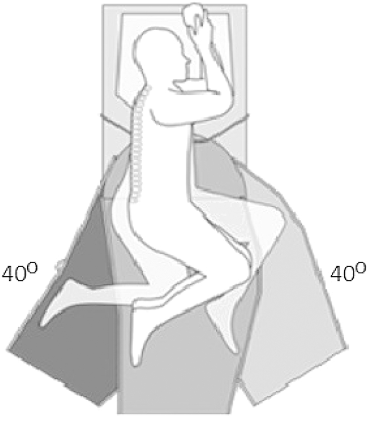
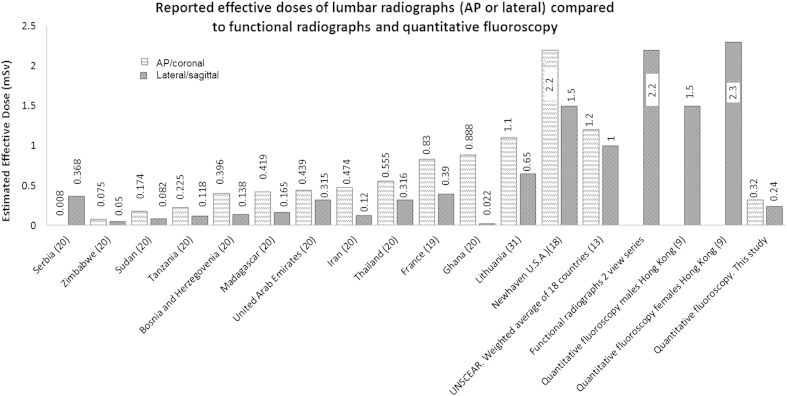
Internationally reported dose area product (DAP) and effective dose data for lumbar spine radiographs were compared with the same for quantitative fluoroscopy and with data from a local hospital for functional radiographs (weight bearing AP, lateral, and/or flexion and extension) ( = 27). The effects of procedure time, age, weight, height and body mass index on the fluoroscopy dose were determined by multiple linear regression using SPSS v19 software (IBM Corp., Armonck, NY, USA).
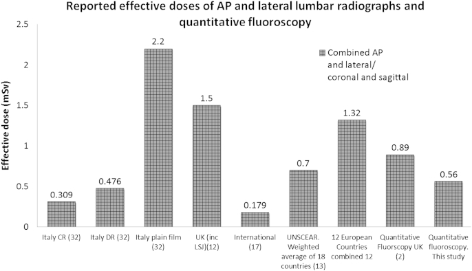
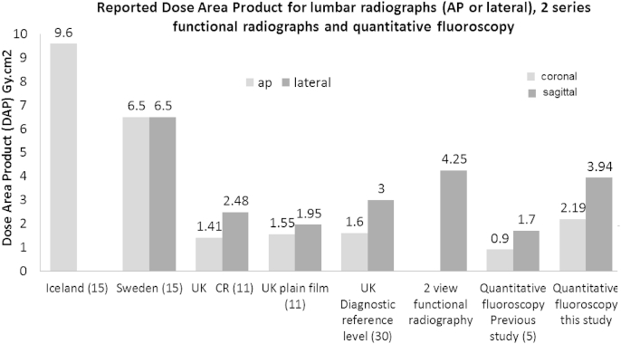
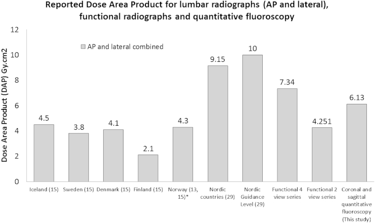
The effective dose (and therefore the estimated risk) for quantitative fluoroscopy is 0.561 mSv which is lower than in most published data for lumbar spine radiography. The dose area product (DAP) for sagittal (flexion + extension) quantitative fluoroscopy is 3.94 Gy cm which is lower than local data for two view (flexion and extension) functional radiographs (4.25 Gy cm), and combined coronal and sagittal dose from quantitative fluoroscopy (6.13 Gy cm) is lower than for four view functional radiography (7.34 Gy cm). Conversely DAP for coronal and sagittal quantitative fluoroscopy combined (6.13 Gy cm) is higher than that published for both lumbar AP or lateral radiographs, with the exception of Nordic countries combined data. Weight, procedure time and age were independently positively associated with total dose, and height (after adjusting for weight) was negatively associated, thus as height increased, the DAP decreased.
定量荧光透视是一种用于评估腰椎连续椎间运动的新兴技术,但关于辐射剂量的信息尚不可用。本研究的目的是比较腰椎定量荧光透视与腰椎X光片的辐射剂量,并确定定量荧光透视中降低剂量的机会。
将国际上报告的腰椎X光片的剂量面积乘积(DAP)和有效剂量数据与定量荧光透视的相同数据以及当地一家医院的功能X光片(负重前后位、侧位和/或屈伸位)(n = 27)的数据进行比较。使用SPSS v19软件(美国纽约州阿蒙克市IBM公司)通过多元线性回归确定检查时间、年龄、体重、身高和体重指数对荧光透视剂量的影响。
定量荧光透视的有效剂量(因此估计风险)为0.561 mSv,低于大多数已发表的腰椎X光摄影数据。矢状面(屈伸)定量荧光透视的剂量面积乘积(DAP)为3.94 Gy·cm,低于双视图(屈伸)功能X光片的当地数据(4.25 Gy·cm),并且定量荧光透视的冠状面和矢状面联合剂量(6.13 Gy·cm)低于四视图功能X光摄影(7.34 Gy·cm)。相反,冠状面和矢状面定量荧光透视联合的DAP(6.13 Gy·cm)高于除北欧国家综合数据外的腰椎前后位或侧位X光片公布的数据。体重、检查时间和年龄与总剂量独立正相关,身高(在调整体重后)与总剂量负相关,因此随着身高增加,DAP降低。