Discipline of Medical Radiation Sciences, School of Molecular and Life Sciences, Curtin University, GPO Box U1987, Perth, Western Australia, 6845, Australia.
School of Health Sciences (HESAV), University of Applied Sciences and Arts Western Switzerland (HES-SO), Av. de Beaumont 21, 1011, Lausanne, Switzerland.
Eur Radiol Exp. 2020 Feb 13;4(1):13. doi: 10.1186/s41747-019-0132-3.
To investigate lateral lumbar spine radiography technical parameters for reduction of effective dose whilst maintaining image quality (IQ).
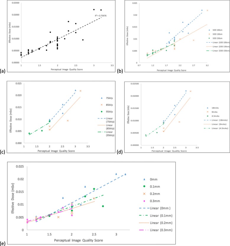
Thirty-six radiograms of an anthropomorphic phantom were acquired using different exposure parameters: source-to-detector distance (SDD) (100, 130 or 150 cm), tube potential (75, 85 or 95 kVp), tube current × exposure time product (4.5, 9, 18 mAs) and additional copper (Cu) filter (no filter, 0.1-, 0.2-, or 0.3-mm thickness. IQ was assessed using an objective approach (contrast-to-noise-ratio [CNR] calculation and magnification measurement) and a perceptual approach (six observers); ED was estimated using the PCXMC 2.0 software. Descriptive statistics, paired t test, and intraclass correlation coefficient (ICC) were used.
The highest ED (0.022 mSv) was found with 100 cm SSD, 75 kVp, 18 mAs, and without Cu filter, whilst the highest CNR (7.23) was achieved at 130 cm SSD, 75 kVp, 18 mAs, and without Cu filter. The lowest ED and CNR were generated at 150 cm SDD, 95 kVp, 4.5 mAs, and 0.3-mm Cu filter. All observers identified the relevant anatomical structures on all images with the lowest ED and IQ. The intra-observer (0.61-0.79) and inter-observer (0.55-0.82) ICC ranged from moderate to excellent.
All relevant anatomical structures were identified on the lateral lumbar spine radiographs despite using low-dose protocols. The lowest ED (0.002 mSv) was obtained with 150 cm SDD, 95 kVp, 4.5 mAs, and 0.3-mm Cu filter. Further technical and clinical studies are needed to verify these preliminary findings.
为了在保持图像质量(IQ)的同时,研究减少侧位腰椎 X 线摄影有效剂量的技术参数。
使用不同的曝光参数对人体模型的 36 张 X 射线照片进行采集:源-探测器距离(SDD)(100、130 或 150cm)、管电压(75、85 或 95kVp)、管电流×曝光时间乘积(4.5、9、18mAs)和附加铜(Cu)滤片(无滤片、0.1、0.2 或 0.3mm 厚)。使用客观方法(对比度噪声比[CNR]计算和放大测量)和主观方法(六位观察者)评估 IQ;使用 PCXMC 2.0 软件估算 ED。使用描述性统计、配对 t 检验和组内相关系数(ICC)进行分析。
在 SDD 为 100cm、管电压为 75kVp、管电流×曝光时间乘积为 18mAs、无 Cu 滤片的情况下,ED 最高(0.022mSv),而在 SDD 为 130cm、管电压为 75kVp、管电流×曝光时间乘积为 18mAs、无 Cu 滤片的情况下,CNR 最高(7.23)。在 SDD 为 150cm、管电压为 95kVp、管电流×曝光时间乘积为 4.5mAs、Cu 滤片为 0.3mm 的情况下,ED 和 CNR 最低。所有观察者均能在 ED 和 IQ 最低的情况下识别出所有相关的解剖结构。观察者内(0.61-0.79)和观察者间(0.55-0.82)ICC 范围为中度至极好。
尽管使用了低剂量方案,但仍能在侧位腰椎 X 射线照片上识别出所有相关的解剖结构。在 SDD 为 150cm、管电压为 95kVp、管电流×曝光时间乘积为 4.5mAs、Cu 滤片为 0.3mm 的情况下,ED 最低(0.002mSv)。需要进一步的技术和临床研究来验证这些初步结果。