Pascarelli Nicola Antonio, Collodel Giulia, Moretti Elena, Cheleschi Sara, Fioravanti Antonella
Department of Medicine, Surgery and Neuroscience, Rheumatology Unit, University of Siena, Policlinico Le Scotte, Viale Bracci 1, Siena 53100, Italy.
Department of Molecular and Developmental Medicine, University of Siena, Siena 53100, Italy.
Int J Mol Sci. 2015 Oct 30;16(11):26019-34. doi: 10.3390/ijms161125936.
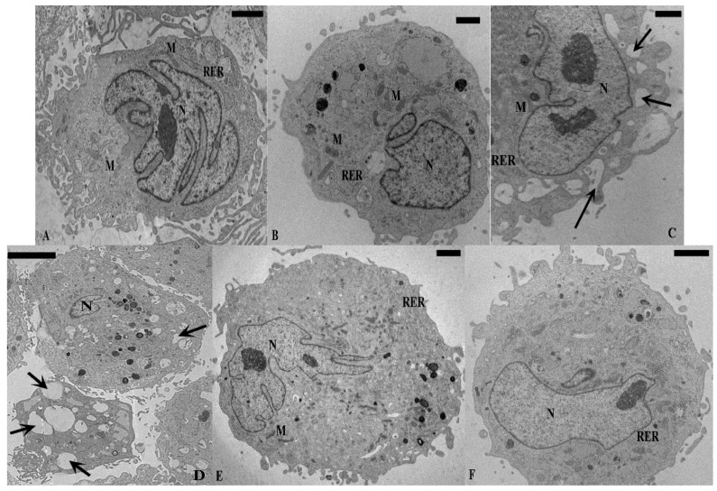
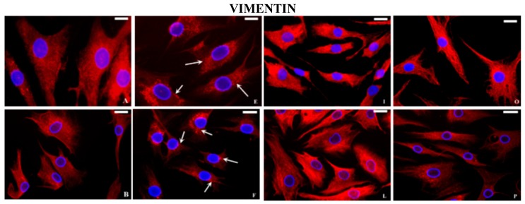
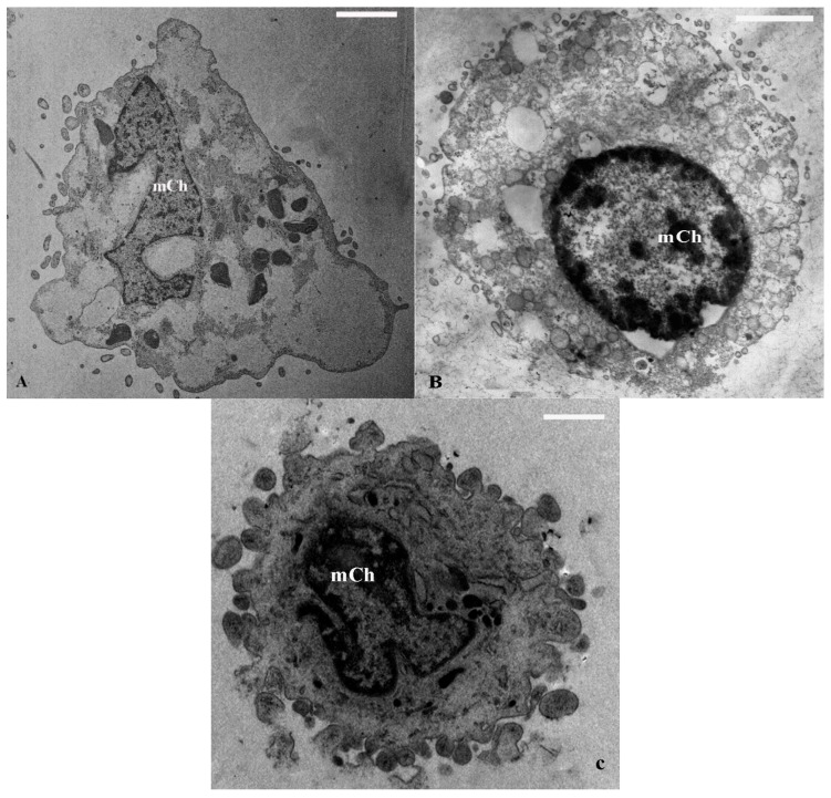
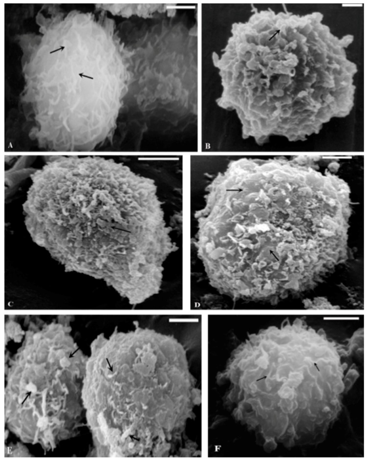
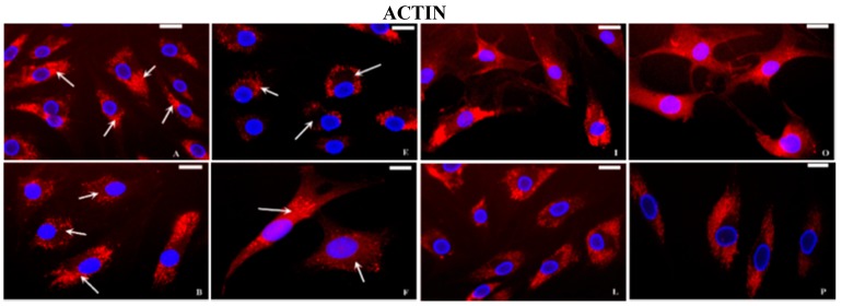
The aim of this study was to examine the ultrastructure and cytoskeletal organization in human normal and Osteoarhritic (OA) chondrocytes, exposed to interleukin-1β (IL-1β) and cyclic hydrostatic pressure (HP). Morphological examination by transmission electron microscopy (TEM) and scanning electron microscopy (SEM) revealed differences between normal and OA chondrocytes at the nuclear and cytoplasmic level. IL-1β (5 ng/mL) induced a decrease of the number of mitochondria and Golgi bodies and a significant increase on the percentage of cells rich in vacuolization and in marginated chromatin. Cyclical HP (1-5 MPa, 0.25 Hz, for 3 h) did not change the morphology of normal chondrocytes, but had a beneficial effect on OA chondrocytes increasing the number of organelles. Normal and OA cells subjected to IL-1β and HP recovered cytoplasmic ultrastructure. Immunofluorescence (IF) examination of normal chondrocytes showed an actin signal polarized on the apical sides of the cytoplasm, tubulin and vimentin uniformly distributed throughout cytoplasm and vinculin revealed a punctuated pattern under the plasma membrane. In OA chondrocytes, these proteins partially lost their organization. Stimulation with IL-1β caused, in both type of cells, modification in the cytoskeletal organization; HP counteracted the negative effects of IL-1β. Our results showed structural differences at nuclear, cytoplasmic and cytoskeletal level between normal and OA chondrocytes. IL-1β induced ultrastructural and cytoskeletal modifications, counteracted by a cyclical low HP.
本研究的目的是检测暴露于白细胞介素-1β(IL-1β)和周期性流体静压力(HP)下的人正常软骨细胞和骨关节炎(OA)软骨细胞的超微结构和细胞骨架组织。通过透射电子显微镜(TEM)和扫描电子显微镜(SEM)进行的形态学检查揭示了正常软骨细胞和OA软骨细胞在细胞核和细胞质水平上的差异。IL-1β(5 ng/mL)导致线粒体和高尔基体数量减少,富含空泡化和边缘化染色质的细胞百分比显著增加。周期性HP(1-5 MPa,0.25 Hz,持续3小时)未改变正常软骨细胞的形态,但对OA软骨细胞具有有益作用,增加了细胞器的数量。暴露于IL-1β和HP的正常和OA细胞恢复了细胞质超微结构。对正常软骨细胞进行的免疫荧光(IF)检查显示,肌动蛋白信号在细胞质的顶端侧极化,微管蛋白和波形蛋白均匀分布于整个细胞质,纽蛋白在质膜下呈现点状分布模式。在OA软骨细胞中,这些蛋白质部分失去了其组织结构。用IL-1β刺激在两种类型的细胞中均导致细胞骨架组织发生改变;HP抵消了IL-1β的负面影响。我们的结果显示正常软骨细胞和OA软骨细胞在细胞核、细胞质和细胞骨架水平上存在结构差异。IL-1β诱导超微结构和细胞骨架改变,而周期性低HP可抵消这些改变。