Han Shuo, Guo Jinhai, Liu Yinan, Zhang Zhi, He Qihua, Li Peng, Zhang Mingzhi, Sun Haojie, Li Ruizhi, Li Yang, Zeng Wotan, Liu Jinwen, Lian Lejian, Gao Yi, Shen Li
Department of Cell Biology, Stem Cell Research Center, Department of Basic Medical Sciences, Peking University Health Science Center, Beijing, People's Republic of China.
Beijing DongFang YaMei Gene Science and Technology Research Institute, Beijing, People's Republic of China.
Oncotarget. 2015 Dec 29;6(42):44452-65. doi: 10.18632/oncotarget.6090.
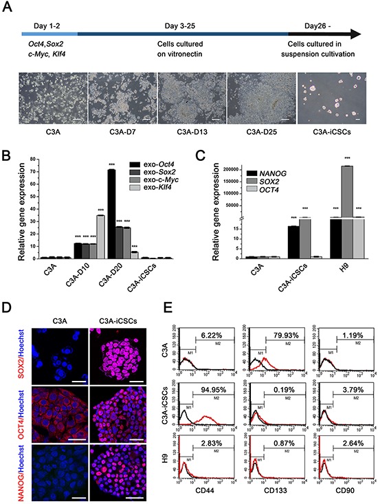
CD44 is a widely known cancer stem cells marker in various cancers and validated to function in tumor growth, survival and tumor metastasis. In this study, we first established C3A-derived liver cancer stem cells by OSKM method [OCT4, SOX2, KLF4, and c-MYC], termed C3A-induced cancer stem cells (C3A-iCSCs) which acquired self-renewal and stemness abilities. Then we found CD44 was positive in C3A-iCSCs and mainly located in cell nuclear. Chromatin immunoprecipitation-quantitative PCR (ChIP-qPCR) results showed nuclear CD44 combined promoter regions of c-MYC and SOX2. These results suggested that CD44 participated in C3A-iCSCs transcriptional regulation. To explore CD44 overall influence in liver cancer stem cells, CD44 was knocked out in C3A-iCSCs using CRISPR/Cas9 technology. Our results showed a dramatic increase in the expression of stem cell markers OCT4, SOX2 and NANOG in CD44- C3A-iCSCs compared with that in CD44+ C3A-iCSCs. Tumor derived from CD44- C3A-iCSCs also displayed well-differentiated tumor cells compared to CD44+ C3A-iCSCs, which suggested CD44- C3A-iCSCs derived tumor cells exhibited lower malignant degree. Our data indicated nuclear CD44 in liver cancer stem cells is responsible for the poorly differentiated highly malignant tumor cells by maintenance of low stemness state.
CD44是一种在多种癌症中广为人知的癌症干细胞标志物,已证实其在肿瘤生长、存活和肿瘤转移中发挥作用。在本研究中,我们首先通过OSKM方法[OCT4、SOX2、KLF4和c-MYC]建立了源自C3A的肝癌干细胞,称为C3A诱导的癌症干细胞(C3A-iCSCs),其获得了自我更新和干性能力。然后我们发现CD44在C3A-iCSCs中呈阳性,且主要位于细胞核中。染色质免疫沉淀-定量PCR(ChIP-qPCR)结果显示,细胞核中的CD44与c-MYC和SOX2的启动子区域结合。这些结果表明CD44参与了C3A-iCSCs的转录调控。为了探究CD44对肝癌干细胞的整体影响,我们使用CRISPR/Cas9技术在C3A-iCSCs中敲除了CD44。我们的结果显示,与CD44+C3A-iCSCs相比,CD44-C3A-iCSCs中干细胞标志物OCT4、SOX2和NANOG的表达显著增加。与CD44+C3A-iCSCs相比,源自CD44-C3A-iCSCs的肿瘤也表现出分化良好的肿瘤细胞,这表明源自CD44-C3A-iCSCs的肿瘤细胞恶性程度较低。我们的数据表明,肝癌干细胞中的细胞核CD44通过维持低干性状态导致低分化的高恶性肿瘤细胞产生。