Dilogo Ismail Hadisoebroto, Kamal Achmad Fauzi, Gunawan Bambang, Rawung Rangga Valentino
Department of Orthopaedic and Traumatology, Cipto Mangunkusumo Hospital, Faculty of Medicine Universitas Indonesia, Jl. Dipenogoro No. 71, Salemba, Jakarta Pusat 10430, Indonesia.
Department of Orthopaedic and Traumatology, Cipto Mangunkusumo Hospital, Faculty of Medicine Universitas Indonesia, Jl. Dipenogoro No. 71, Salemba, Jakarta Pusat 10430, Indonesia.
Int J Surg Case Rep. 2015;17:106-11. doi: 10.1016/j.ijscr.2015.10.040. Epub 2015 Nov 5.
Osteofibrous dysplasia is a rare non-neoplastic disease that is almost exclusive to pediatric tibial diaphysis. Wide excision of the lesion is recommended to avoid recurrence. However, such radical surgery will results in large segmental bone defects that will require further extensive reconstructive surgery. We report a novel approach of treating bone defect by implementing the diamond concept of bone healing using autologous bone marrow derived mesenchymal stem cells (BM-MSCs).
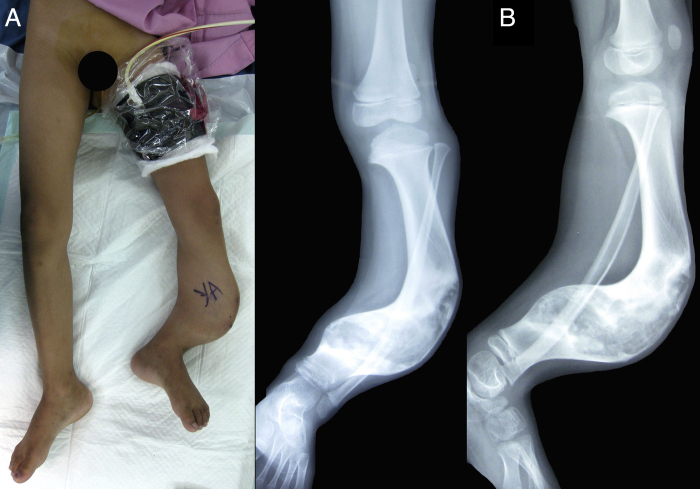
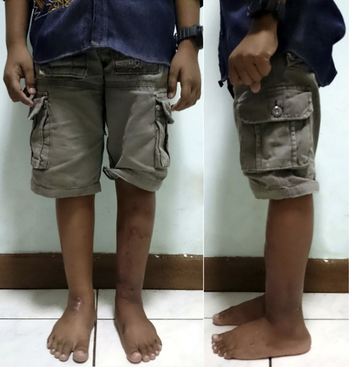
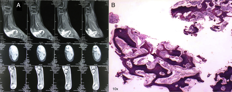
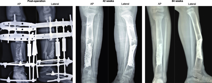
An eight-year-old Indonesian male presented with severe bowing deformity of the left lower leg. Radiographic and histological analysis confirmed the diagnosis of osteofibrous dysplasia. A wide excision of the defect was made leaving a critical-sized bone defect. A combination of autologous transplantation of 50 million BM-MSCs, hydroxyapatite (HA) granules, bone morphogenic protein 2 (BMP-2) and Djoko-Zarov hybrid circular external fixator was used to treat the defect. The outcomes measured were subjective complaints, functionality based on LEFS and radiological assessments.
Radiographic assessments showed successful new bone tissue formation and integration of implanted HA granules. The external fixator was removed at 42 weeks after adequate callus formation and clinical stability was achieved. The patient underwent progressive functional improvements and reached a near normal functionality of 90% LEFS at 84 week. No therapy side effect or complication was reported.
Osteofibrous dysplasia was successfully excised without signs of recurrence after 84-week follow-up. Autologous transplantation of augmented BM-MSCs has successfully created new normal bone tissue without causing any side effect and had significantly improved the patient's quality of life.
骨纤维发育不良是一种罕见的非肿瘤性疾病,几乎仅见于小儿胫骨干。建议广泛切除病灶以避免复发。然而,这种根治性手术会导致大段骨缺损,需要进一步进行广泛的重建手术。我们报告一种采用自体骨髓间充质干细胞(BM-MSCs)运用骨愈合的钻石概念治疗骨缺损的新方法。
一名8岁印度尼西亚男性,左小腿严重弓形畸形。影像学和组织学分析确诊为骨纤维发育不良。广泛切除缺损部位,留下临界大小的骨缺损。采用5000万BM-MSCs自体移植、羟基磷灰石(HA)颗粒、骨形态发生蛋白2(BMP-2)和佐科-扎罗夫混合环形外固定器联合治疗该缺损。测量的结果包括主观症状、基于下肢功能评分(LEFS)的功能以及影像学评估。
影像学评估显示成功形成了新的骨组织并植入的HA颗粒融合。在骨痂形成充分且达到临床稳定后,于42周拆除外固定器。患者功能逐渐改善,在84周时达到近正常功能,LEFS为90%。未报告治疗副作用或并发症。
骨纤维发育不良在84周随访后成功切除,无复发迹象。增强型BM-MSCs自体移植成功创建了新的正常骨组织,未产生任何副作用,并显著改善了患者的生活质量。