Motley Alison M, Galvin Paul C, Ekal Lakhan, Nuttall James M, Hettema Ewald H
Department of Molecular Biology and Biotechnology, University of Sheffield, Sheffield S10 2TN, England, UK.
Department of Molecular Biology and Biotechnology, University of Sheffield, Sheffield S10 2TN, England, UK
J Cell Biol. 2015 Dec 7;211(5):1041-56. doi: 10.1083/jcb.201412066.
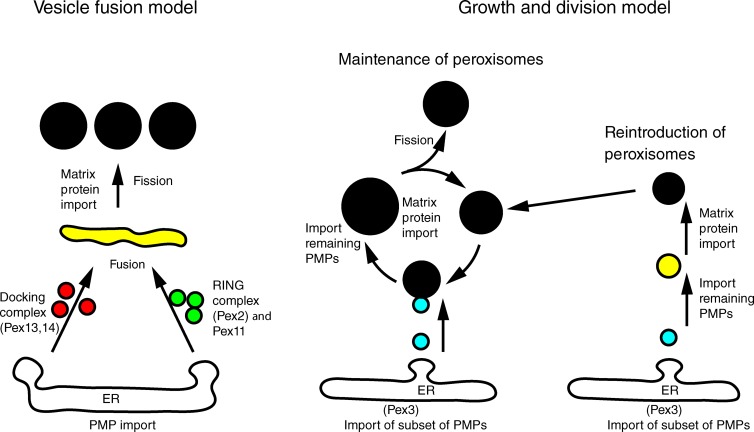
A recent model for peroxisome biogenesis postulates that peroxisomes form de novo continuously in wild-type cells by heterotypic fusion of endoplasmic reticulum-derived vesicles containing distinct sets of peroxisomal membrane proteins. This model proposes a role in vesicle fusion for the Pex1/Pex6 complex, which has an established role in matrix protein import. The growth and division model proposes that peroxisomes derive from existing peroxisomes. We tested these models by reexamining the role of Pex1/Pex6 and dynamin-related proteins in peroxisome biogenesis. We found that induced depletion of Pex1 blocks the import of matrix proteins but does not affect membrane protein delivery to peroxisomes; markers for the previously reported distinct vesicles colocalize in pex1 and pex6 cells; peroxisomes undergo continued growth if fission is blocked. Our data are compatible with the established primary role of the Pex1/Pex6 complex in matrix protein import and show that peroxisomes in Saccharomyces cerevisiae multiply mainly by growth and division.
最近的过氧化物酶体生物发生模型假定,在野生型细胞中,过氧化物酶体通过内质网衍生的、含有不同过氧化物酶体膜蛋白组的囊泡的异型融合而持续从头形成。该模型提出Pex1/Pex6复合体在囊泡融合中起作用,而该复合体在基质蛋白输入中已有明确作用。生长和分裂模型提出过氧化物酶体源自现有的过氧化物酶体。我们通过重新审视Pex1/Pex6和动力蛋白相关蛋白在过氧化物酶体生物发生中的作用来测试这些模型。我们发现,诱导性地耗尽Pex1会阻断基质蛋白的输入,但不影响膜蛋白向过氧化物酶体的递送;先前报道的不同囊泡的标记物在pex1和pex6细胞中共定位;如果裂变受阻,过氧化物酶体会持续生长。我们的数据与Pex1/Pex6复合体在基质蛋白输入中的既定主要作用相符,并表明酿酒酵母中的过氧化物酶体主要通过生长和分裂进行增殖。