Saenz Daniel L, Yan Yue, Christensen Neil, Henzler Margaret A, Forrest Lisa J, Bayouth John E, Paliwal Bhudatt R
University of Wisconsin-Madison.
J Appl Clin Med Phys. 2015 Nov 8;16(6):30-40. doi: 10.1120/jacmp.v16i6.5353.
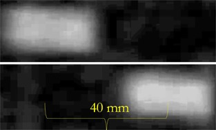
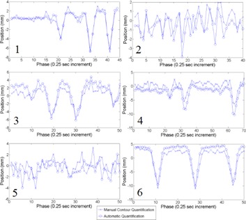
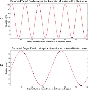
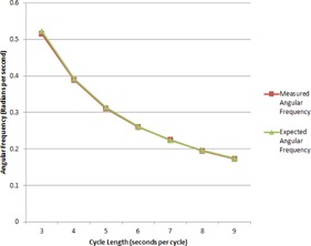
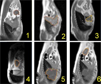
ViewRay is a novel MR-guided radiotherapy system capable of imaging in near real-time at four frames per second during treatment using 0.35T field strength. It allows for improved gating techniques and adaptive radiotherapy. Three cobalt-60 sources (~ 15,000 Curies) permit multiple-beam, intensity-modulated radiation therapy. The primary aim of this study is to assess the imaging stability, accuracy, and automatic segmentation algorithm capability to track motion in simulated and in vivo targets. Magnetic resonance imaging (MRI) characteristics of the system were assessed using the American College of Radiology (ACR)-recommended phantom and accreditation protocol. Images of the ACR phantom were acquired using a head coil following the ACR scanning instructions. ACR recommended T1- and T2-weighted sequences were evaluated. Nine measurements were performed over a period of seven months, on just over a monthly basis, to establish consistency. A silicon dielectric gel target was attached to the motor via a rod. 40 mm total amplitude was used with cycles of 3 to 9 s in length in a sinusoidal trajectory. Trajectories of six moving clinical targets in four canine patients were quantified and tracked. ACR phantom images were analyzed, and the results were compared with the ACR acceptance levels. Measured slice thickness accuracies were within the acceptance limits. In the 0.35 T system, the image intensity uniformity was also within the ACR acceptance limit. Over the range of cycle lengths, representing a wide range of breathing rates in patients imaged at four frames/s, excellent agreement was observed between the expected and measured target trajectories. In vivo canine targets, including the gross target volume (GTV), as well as other abdominal soft tissue structures, were visualized with inherent MR contrast, allowing for preliminary results of target tracking.
ViewRay是一种新型的磁共振引导放射治疗系统,在使用0.35T场强进行治疗期间,能够以每秒4帧的速度进行近实时成像。它允许改进门控技术和自适应放射治疗。三个钴 - 60源(约15,000居里)可实现多束强度调制放射治疗。本研究的主要目的是评估成像稳定性、准确性以及自动分割算法在模拟和体内目标中跟踪运动的能力。使用美国放射学会(ACR)推荐的体模和认证方案评估该系统的磁共振成像(MRI)特性。按照ACR扫描说明,使用头部线圈采集ACR体模的图像。对ACR推荐的T1加权和T2加权序列进行评估。在七个月的时间内进行了九次测量,大约每月一次,以确定一致性。通过一根杆将硅介电凝胶靶连接到电机上。在正弦轨迹中使用40毫米的总振幅,周期长度为3至9秒。对四只犬类患者体内六个移动临床目标的轨迹进行了量化和跟踪。分析了ACR体模图像,并将结果与ACR验收标准进行了比较。测量的切片厚度精度在验收范围内。在0.35T系统中,图像强度均匀性也在ACR验收范围内。在代表以每秒4帧成像的患者的广泛呼吸频率的周期长度范围内,观察到预期和测量的目标轨迹之间具有极好的一致性。通过固有的磁共振对比度可以可视化体内犬类目标,包括大体肿瘤体积(GTV)以及其他腹部软组织结构,从而获得目标跟踪的初步结果。