• 文献检索
  • 文档翻译
  • 深度研究
  • 学术资讯
  • Suppr Zotero 插件Zotero 插件
  • 邀请有礼
  • 套餐&价格
  • 历史记录
应用&插件
Suppr Zotero 插件Zotero 插件浏览器插件Mac 客户端Windows 客户端微信小程序
定价
高级版会员购买积分包购买API积分包
服务
文献检索文档翻译深度研究API 文档MCP 服务
关于我们
关于 Suppr公司介绍联系我们用户协议隐私条款
关注我们

Suppr 超能文献

核心技术专利:CN118964589B侵权必究
粤ICP备2023148730 号-1Suppr @ 2026

文献检索

告别复杂PubMed语法,用中文像聊天一样搜索,搜遍4000万医学文献。AI智能推荐,让科研检索更轻松。

立即免费搜索

文件翻译

保留排版,准确专业,支持PDF/Word/PPT等文件格式,支持 12+语言互译。

免费翻译文档

深度研究

AI帮你快速写综述,25分钟生成高质量综述,智能提取关键信息,辅助科研写作。

立即免费体验

基于模糊逻辑的神经元荧光显微镜图像中连接点和末端的检测与特征提取。

Fuzzy-Logic Based Detection and Characterization of Junctions and Terminations in Fluorescence Microscopy Images of Neurons.

机构信息

Biomedical Imaging Group Rotterdam, Departments of Medical Informatics and Radiology, Erasmus University Medical Center, Rotterdam, the Netherlands.

出版信息

Neuroinformatics. 2016 Apr;14(2):201-19. doi: 10.1007/s12021-015-9287-0.

DOI:10.1007/s12021-015-9287-0
PMID:26701809
原文链接:https://pmc.ncbi.nlm.nih.gov/articles/PMC4823367/
Abstract

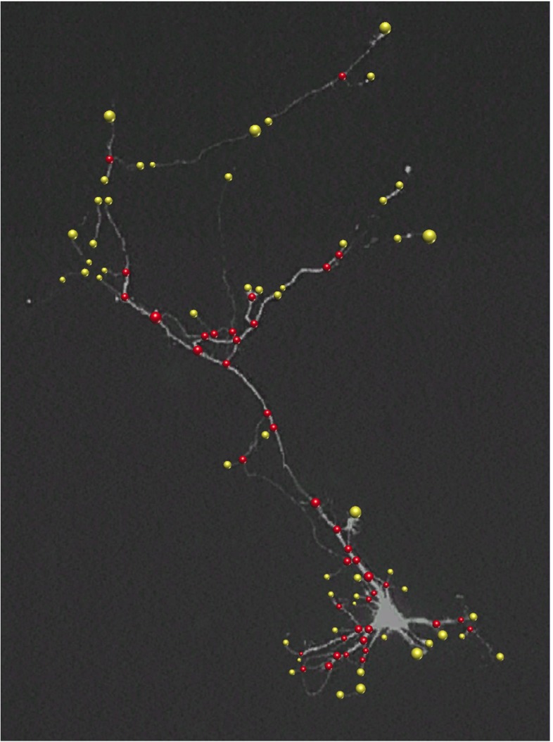
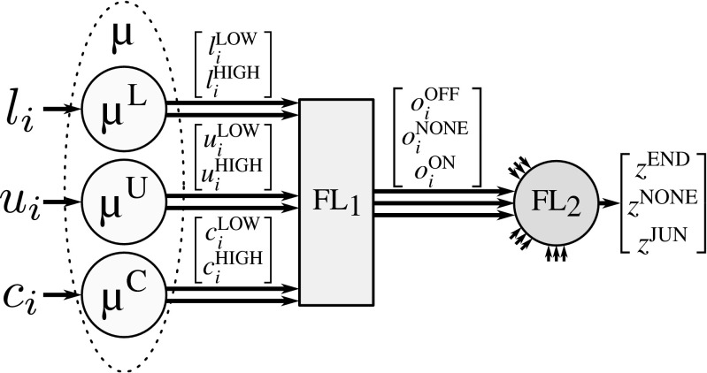
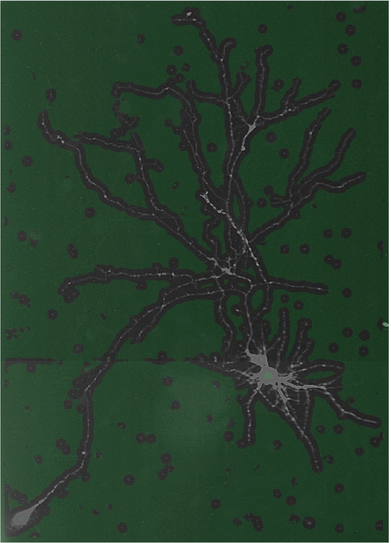
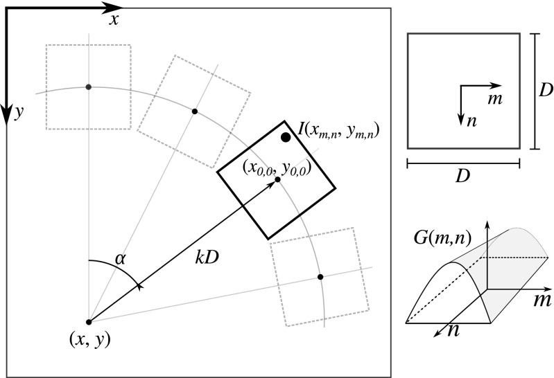
Digital reconstruction of neuronal cell morphology is an important step toward understanding the functionality of neuronal networks. Neurons are tree-like structures whose description depends critically on the junctions and terminations, collectively called critical points, making the correct localization and identification of these points a crucial task in the reconstruction process. Here we present a fully automatic method for the integrated detection and characterization of both types of critical points in fluorescence microscopy images of neurons. In view of the majority of our current studies, which are based on cultured neurons, we describe and evaluate the method for application to two-dimensional (2D) images. The method relies on directional filtering and angular profile analysis to extract essential features about the main streamlines at any location in an image, and employs fuzzy logic with carefully designed rules to reason about the feature values in order to make well-informed decisions about the presence of a critical point and its type. Experiments on simulated as well as real images of neurons demonstrate the detection performance of our method. A comparison with the output of two existing neuron reconstruction methods reveals that our method achieves substantially higher detection rates and could provide beneficial information to the reconstruction process.

摘要

神经元细胞形态的数字重建是理解神经元网络功能的重要步骤。神经元是树状结构,其描述取决于连接点和端点,统称为关键点,因此正确定位和识别这些点是重建过程中的关键任务。在这里,我们提出了一种用于荧光显微镜神经元图像中两种类型关键点的集成检测和特征描述的全自动方法。鉴于我们目前的大多数研究都是基于培养的神经元,我们将描述和评估该方法在二维(2D)图像中的应用。该方法依赖于方向滤波和角度轮廓分析来提取图像中任何位置的主要流线的基本特征,并使用具有精心设计规则的模糊逻辑来推理特征值,以便对关键点的存在及其类型做出明智的决策。对神经元的模拟和真实图像的实验证明了我们方法的检测性能。与两种现有神经元重建方法的输出结果进行比较表明,我们的方法可以实现更高的检测率,并为重建过程提供有益的信息。

https://cdn.ncbi.nlm.nih.gov/pmc/blobs/e47b/4823367/0d38e38b8863/12021_2015_9287_Fig7_HTML.jpg
https://cdn.ncbi.nlm.nih.gov/pmc/blobs/e47b/4823367/cd4e7037ac02/12021_2015_9287_Fig1_HTML.jpg
https://cdn.ncbi.nlm.nih.gov/pmc/blobs/e47b/4823367/f2f1dfce7989/12021_2015_9287_Fig2_HTML.jpg
https://cdn.ncbi.nlm.nih.gov/pmc/blobs/e47b/4823367/093b37204c88/12021_2015_9287_Fig3_HTML.jpg
https://cdn.ncbi.nlm.nih.gov/pmc/blobs/e47b/4823367/0d38e38b8863/12021_2015_9287_Fig7_HTML.jpg

相似文献

1
Fuzzy-Logic Based Detection and Characterization of Junctions and Terminations in Fluorescence Microscopy Images of Neurons.基于模糊逻辑的神经元荧光显微镜图像中连接点和末端的检测与特征提取。
Neuroinformatics. 2016 Apr;14(2):201-19. doi: 10.1007/s12021-015-9287-0.
2
Improved detection of soma location and morphology in fluorescence microscopy images of neurons.在神经元荧光显微镜图像中对胞体位置和形态的检测得到改进。
J Neurosci Methods. 2016 Dec 1;274:61-70. doi: 10.1016/j.jneumeth.2016.09.007. Epub 2016 Sep 26.
3
A Multiscale Ray-Shooting Model for Termination Detection of Tree-Like Structures in Biomedical Images.一种用于生物医学图像中树状结构终点检测的多尺度光线投射模型。
IEEE Trans Med Imaging. 2019 Aug;38(8):1923-1934. doi: 10.1109/TMI.2019.2893117. Epub 2019 Jan 15.
4
Fuzzy Index to Evaluate Edge Detection in Digital Images.用于评估数字图像边缘检测的模糊指标。
PLoS One. 2015 Jun 26;10(6):e0131161. doi: 10.1371/journal.pone.0131161. eCollection 2015.
5
Automatic thoracic anatomy segmentation on CT images using hierarchical fuzzy models and registration.使用分层模糊模型和配准技术对CT图像进行胸部解剖结构自动分割
Med Phys. 2016 Mar;43(3):1487-500. doi: 10.1118/1.4942486.
6
Spherical-Patches Extraction for Deep-Learning-Based Critical Points Detection in 3D Neuron Microscopy Images.基于球面补丁提取的三维神经元显微镜图像中关键点检测的深度学习方法。
IEEE Trans Med Imaging. 2021 Feb;40(2):527-538. doi: 10.1109/TMI.2020.3031289. Epub 2021 Feb 2.
7
Automated Neuron Reconstruction from 3D Fluorescence Microscopy Images Using Sequential Monte Carlo Estimation.基于序贯蒙特卡罗估计的 3D 荧光显微镜图像自动神经元重建。
Neuroinformatics. 2019 Jul;17(3):423-442. doi: 10.1007/s12021-018-9407-8.
8
Quantitative Analysis of Rat Dorsal Root Ganglion Neurons Cultured on Microelectrode Arrays Based on Fluorescence Microscopy Image Processing.基于荧光显微镜图像处理的大鼠背根神经节神经元在微电极阵列上的定量分析。
Int J Neural Syst. 2015 Dec;25(8):1550033. doi: 10.1142/S0129065715500331. Epub 2015 Sep 14.
9
Spatial fuzzy c-means algorithm with adaptive fuzzy exponent selection for robust vermilion border detection in healthy and diseased lower lips.用于健康和患病下唇的稳健唇红线检测的具有自适应模糊指数选择的空间模糊 c-均值算法。
Comput Methods Programs Biomed. 2014 May;114(3):291-301. doi: 10.1016/j.cmpb.2014.02.017. Epub 2014 Mar 6.
10
Extracellular spike detection from multiple electrode array using novel intelligent filter and ensemble fuzzy decision making.使用新型智能滤波器和集成模糊决策从多电极阵列检测细胞外尖峰。
J Neurosci Methods. 2015 Jan 15;239:129-38. doi: 10.1016/j.jneumeth.2014.10.006. Epub 2014 Oct 18.

引用本文的文献

1
DNeuroMAT: A Deep-Learning-Based Neuron Morphology Analysis Toolbox.DNeuroMAT:基于深度学习的神经元形态分析工具箱。
Methods Mol Biol. 2024;2831:179-197. doi: 10.1007/978-1-0716-3969-6_12.
2
Brain-Wide Shape Reconstruction of a Traced Neuron Using the Convex Image Segmentation Method.利用凸图像分割方法进行追踪神经元的全脑形态重建。
Neuroinformatics. 2020 Apr;18(2):199-218. doi: 10.1007/s12021-019-09434-x.
3
Optimization of Traced Neuron Skeleton Using Lasso-Based Model.基于套索模型的追踪神经元骨架优化

本文引用的文献

1
TReMAP: Automatic 3D Neuron Reconstruction Based on Tracing, Reverse Mapping and Assembling of 2D Projections.TReMAP:基于二维投影的追踪、反向映射和组装的自动三维神经元重建
Neuroinformatics. 2016 Jan;14(1):41-50. doi: 10.1007/s12021-015-9278-1.
2
Automatic Morphological Reconstruction of Neurons from Multiphoton and Confocal Microscopy Images Using 3D Tubular Models.使用3D管状模型从多光子和共聚焦显微镜图像自动重建神经元形态
Neuroinformatics. 2015 Jul;13(3):297-320. doi: 10.1007/s12021-014-9253-2.
3
Dendritic structural degeneration is functionally linked to cellular hyperexcitability in a mouse model of Alzheimer's disease.
Front Neuroanat. 2019 Feb 21;13:18. doi: 10.3389/fnana.2019.00018. eCollection 2019.
4
Automated Neuron Reconstruction from 3D Fluorescence Microscopy Images Using Sequential Monte Carlo Estimation.基于序贯蒙特卡罗估计的 3D 荧光显微镜图像自动神经元重建。
Neuroinformatics. 2019 Jul;17(3):423-442. doi: 10.1007/s12021-018-9407-8.
5
Cooperative carotid artery centerline extraction in MRI.磁共振成像中的协同颈动脉中心线提取。
PLoS One. 2018 May 30;13(5):e0197180. doi: 10.1371/journal.pone.0197180. eCollection 2018.
树突结构退化与阿尔茨海默病小鼠模型中的细胞过度兴奋在功能上相关联。
Neuron. 2014 Dec 3;84(5):1023-33. doi: 10.1016/j.neuron.2014.10.024. Epub 2014 Nov 13.
4
Objective comparison of particle tracking methods.目的比较粒子跟踪方法。
Nat Methods. 2014 Mar;11(3):281-9. doi: 10.1038/nmeth.2808. Epub 2014 Jan 19.
5
Extensible visualization and analysis for multidimensional images using Vaa3D.使用 Vaa3D 进行多维图像的可扩展可视化和分析。
Nat Protoc. 2014 Jan;9(1):193-208. doi: 10.1038/nprot.2014.011. Epub 2014 Jan 2.
6
The Filament Editor: an interactive software environment for visualization, proof-editing and analysis of 3D neuron morphology.《丝状编辑软件:用于 3D 神经元形态可视化、校对编辑和分析的交互软件环境》。
Neuroinformatics. 2014 Apr;12(2):325-39. doi: 10.1007/s12021-013-9213-2.
7
The control of branching morphogenesis.分支形态发生的控制。
Open Biol. 2013 Sep 4;3(9):130088. doi: 10.1098/rsob.130088.
8
APP2: automatic tracing of 3D neuron morphology based on hierarchical pruning of a gray-weighted image distance-tree.APP2:基于灰度加权图像距离树的层次剪枝的 3D 神经元形态自动追踪。
Bioinformatics. 2013 Jun 1;29(11):1448-54. doi: 10.1093/bioinformatics/btt170. Epub 2013 Apr 19.
9
NIH Image to ImageJ: 25 years of image analysis.NIH 图像到 ImageJ:25 年的图像分析。
Nat Methods. 2012 Jul;9(7):671-5. doi: 10.1038/nmeth.2089.
10
Segmentation and tracing of single neurons from 3D confocal microscope images.从三维共聚焦显微镜图像中分割和追踪单个神经元。
IEEE J Biomed Health Inform. 2013 Mar;17(2):319-35. doi: 10.1109/TITB.2012.2209670. Epub 2012 Jul 20.