Zhang Haifei, Cannell Mark B, Kim Shang Jin, Watson Judy J, Norman Ruth, Calaghan Sarah C, Orchard Clive H, James Andrew F
Cardiovascular Research Laboratories, School of Physiology, Pharmacology & Neuroscience, University of Bristol, Bristol, BS8 1TD, United Kingdom.
School of Biomedical Sciences, Garstang, University of Leeds, Leeds, LS2 9JT, United Kingdom.
PLoS One. 2015 Dec 29;10(12):e0144309. doi: 10.1371/journal.pone.0144309. eCollection 2015.
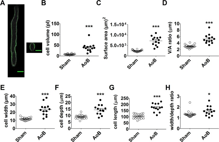
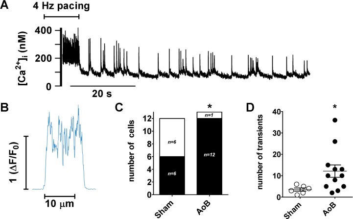
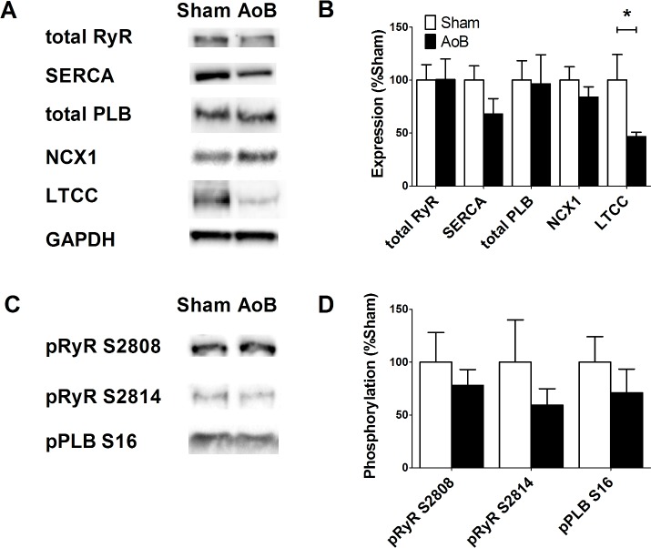
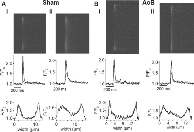
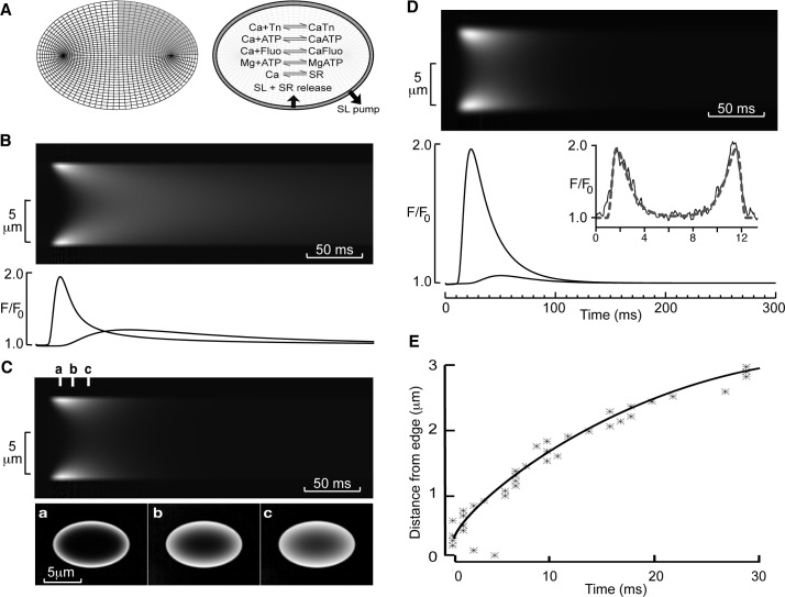
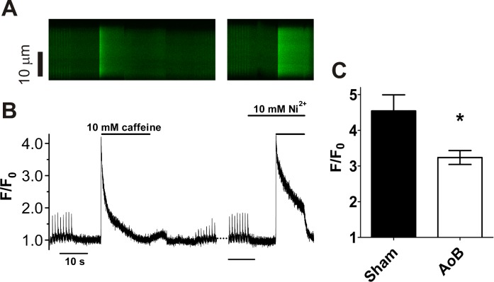
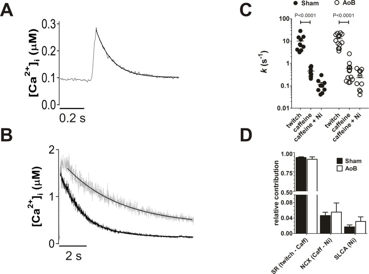
Atrial remodeling due to elevated arterial pressure predisposes the heart to atrial fibrillation (AF). Although abnormal sarcoplasmic reticulum (SR) function has been associated with AF, there is little information on the effects of elevated afterload on atrial Ca2+-handling. We investigated the effects of ascending aortic banding (AoB) on Ca2+-handling in rat isolated atrial myocytes in comparison to age-matched sham-operated animals (Sham). Myocytes were either labelled for ryanodine receptor (RyR) or loaded with fluo-3-AM and imaged by confocal microscopy. AoB myocytes were hypertrophied in comparison to Sham controls (P<0.0001). RyR labeling was localized to the z-lines and to the cell edge. There were no differences between AoB and Sham in the intensity or pattern of RyR-staining. In both AoB and Sham, electrical stimulation evoked robust SR Ca2+-release at the cell edge whereas Ca2+ transients at the cell center were much smaller. Western blotting showed a decreased L-type Ca channel expression but no significant changes in RyR or RyR phosphorylation or in expression of Na+/Ca2+ exchanger, SR Ca2+ ATPase or phospholamban. Mathematical modeling indicated that [Ca2+]i transients at the cell center were accounted for by simple centripetal diffusion of Ca2+ released at the cell edge. In contrast, caffeine (10 mM) induced Ca2+ release was uniform across the cell. The caffeine-induced transient was smaller in AoB than in Sham, suggesting a reduced SR Ca2+-load in hypertrophied cells. There were no significant differences between AoB and Sham cells in the rate of Ca2+ extrusion during recovery of electrically-stimulated or caffeine-induced transients. The incidence and frequency of spontaneous Ca2+-transients following rapid-pacing (4 Hz) was greater in AoB than in Sham myocytes. In conclusion, elevated afterload causes cellular hypertrophy and remodeling of atrial SR Ca2+-release.
动脉血压升高引起的心房重构使心脏易患心房颤动(AF)。尽管异常的肌浆网(SR)功能与AF有关,但关于后负荷升高对心房Ca2+处理的影响的信息很少。我们研究了升主动脉缩窄(AoB)对大鼠离体心房肌细胞Ca2+处理的影响,并与年龄匹配的假手术动物(Sham)进行比较。将肌细胞用ryanodine受体(RyR)标记或用fluo-3-AM加载,并用共聚焦显微镜成像。与Sham对照组相比,AoB肌细胞肥大(P<0.0001)。RyR标记定位于z线和细胞边缘。AoB组和Sham组在RyR染色的强度或模式上没有差异。在AoB组和Sham组中,电刺激均在细胞边缘引起强烈的SR Ca2+释放,而细胞中心的Ca2+瞬变则小得多。蛋白质印迹显示L型钙通道表达降低,但RyR或RyR磷酸化以及钠/钙交换体、SR Ca2+ ATP酶或受磷蛋白的表达没有显著变化。数学模型表明,细胞中心的[Ca2+]i瞬变是由细胞边缘释放的Ca2+的简单向心扩散引起的。相反,咖啡因(10 mM)诱导的Ca2+释放在整个细胞中是均匀的。咖啡因诱导的瞬变在AoB组中比在Sham组中小,表明肥大细胞中SR Ca2+负荷降低。在电刺激或咖啡因诱导的瞬变恢复过程中,AoB组和Sham组细胞的Ca2+外排速率没有显著差异。快速起搏(4 Hz)后自发Ca2+瞬变的发生率和频率在AoB组中比在Sham组心肌细胞中更高。总之,后负荷升高导致细胞肥大和心房SR Ca2+释放的重构。