• 文献检索
  • 文档翻译
  • 深度研究
  • 学术资讯
  • Suppr Zotero 插件Zotero 插件
  • 邀请有礼
  • 套餐&价格
  • 历史记录
应用&插件
Suppr Zotero 插件Zotero 插件浏览器插件Mac 客户端Windows 客户端微信小程序
定价
高级版会员购买积分包购买API积分包
服务
文献检索文档翻译深度研究API 文档MCP 服务
关于我们
关于 Suppr公司介绍联系我们用户协议隐私条款
关注我们

Suppr 超能文献

核心技术专利:CN118964589B侵权必究
粤ICP备2023148730 号-1Suppr @ 2026

文献检索

告别复杂PubMed语法,用中文像聊天一样搜索,搜遍4000万医学文献。AI智能推荐,让科研检索更轻松。

立即免费搜索

文件翻译

保留排版,准确专业,支持PDF/Word/PPT等文件格式,支持 12+语言互译。

免费翻译文档

深度研究

AI帮你快速写综述,25分钟生成高质量综述,智能提取关键信息,辅助科研写作。

立即免费体验

兔和鼠心肌细胞 T 管系统中的溶质运动。

Solute movement in the t-tubule system of rabbit and mouse cardiomyocytes.

机构信息

School of Physiology, Pharmacology & Neuroscience, Faculty of Biomedical Sciences, University of Bristol, BS8 1TD Bristol, United Kingdom.

Institute for Experimental Cardiovascular Medicine, University Heart Centre Freiburg - Bad Krozingen, Faculty of Medicine, University of Freiburg, 79110 Freiburg, Germany.

出版信息

Proc Natl Acad Sci U S A. 2018 Jul 24;115(30):E7073-E7080. doi: 10.1073/pnas.1805979115. Epub 2018 Jul 10.

DOI:10.1073/pnas.1805979115
PMID:29991602
原文链接:https://pmc.ncbi.nlm.nih.gov/articles/PMC6065001/
Abstract

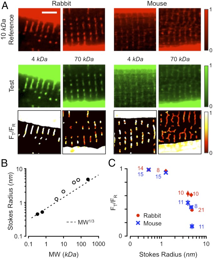
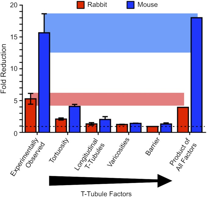
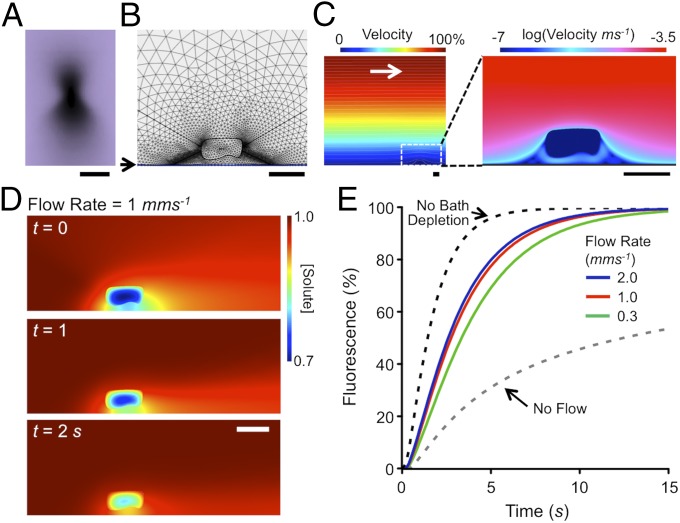
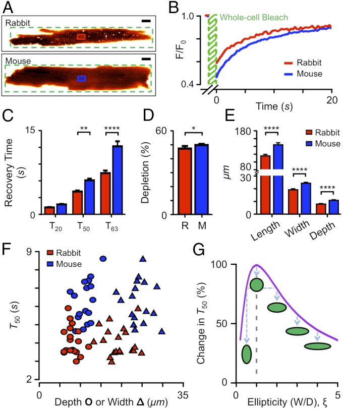
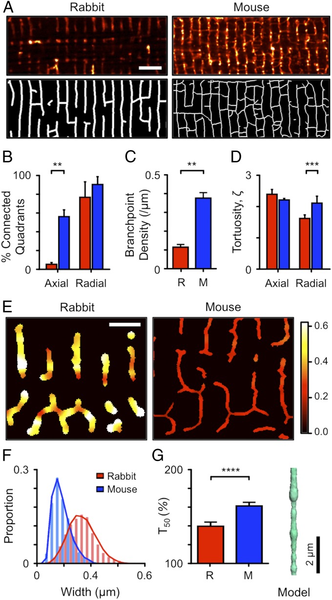
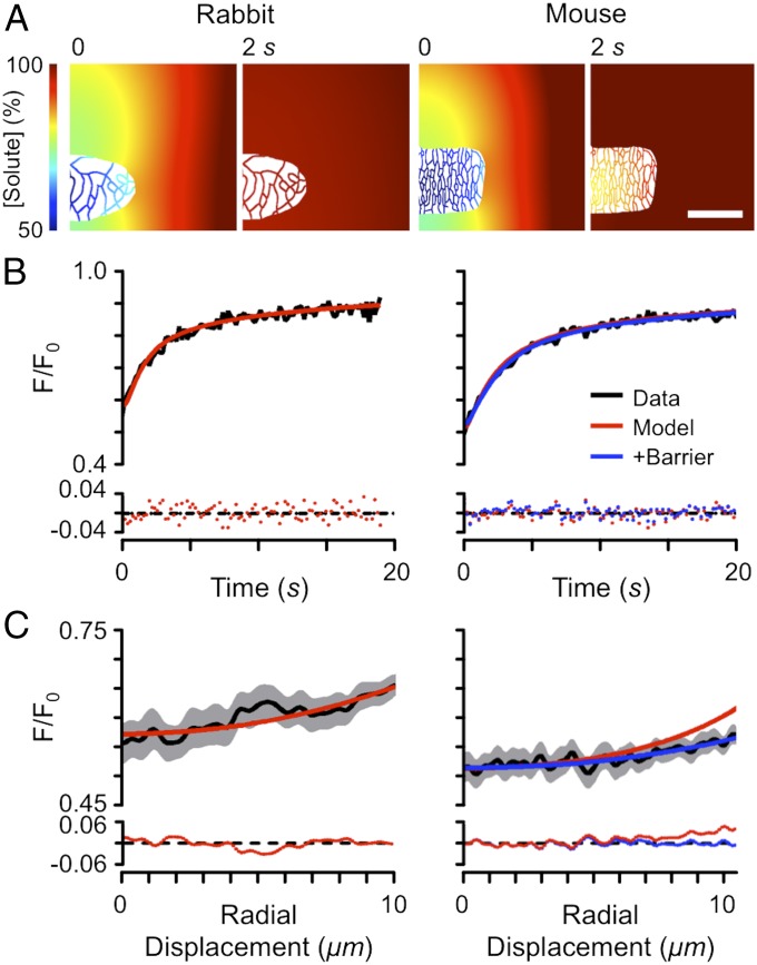
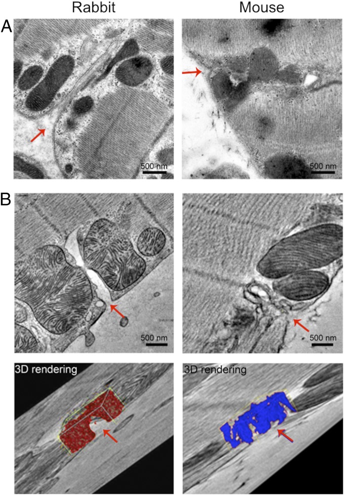
Cardiac transverse (t-) tubules carry both electrical excitation and solutes toward the cell center but their ability to transport small molecules is unclear. While fluorescence recovery after photobleaching (FRAP) can provide an approach to measure local solute movement, extraction of diffusion coefficients is confounded by cell and illumination beam geometries. In this study, we use measured cellular geometry and detailed computer modeling to derive the apparent diffusion coefficient of a 1-kDa solute inside the t-tubular system of rabbit and mouse ventricular cardiomyocytes. This approach shows that diffusion within individual t-tubules is more rapid than previously reported. T-tubule tortuosity, varicosities, and the presence of longitudinal elements combine to substantially reduce the apparent rate of solute movement. In steady state, large (>4 kDa) solutes did not freely fill the t-tubule lumen of both species and <50% of the t-tubule volume was available to solutes >70 kDa. Detailed model fitting of FRAP data suggests that solute diffusion is additionally restricted at the t-tubular entrance and this effect was larger in mouse than in rabbit. The possible structural basis of this effect was investigated using electron microscopy and tomography. Near the cell surface, mouse t-tubules are more tortuous and filled with an electron-dense ground substance, previously identified as glycocalyx and a polyanionic mesh. Solute movement in the t-tubule network of rabbit and mouse appears to be explained by their different geometric properties, which impacts the use of these species for understanding t-tubule function and the consequences of changes associated with t-tubule disease.

摘要

心的横向(t-)小管携带电兴奋和溶质向细胞中心,但它们运输小分子的能力尚不清楚。虽然光漂白后荧光恢复(FRAP)可以提供一种测量局部溶质运动的方法,但扩散系数的提取受到细胞和照明光束几何形状的影响。在这项研究中,我们使用测量的细胞几何形状和详细的计算机建模来推导兔和鼠心室心肌细胞 t-小管系统内 1kDa 溶质的表观扩散系数。这种方法表明,单个 t-小管内的扩散速度比以前报道的要快。t-小管的扭曲、静脉曲张和纵向元素的存在结合起来,大大降低了溶质运动的表观速率。在稳态下,大于 4kDa 的大溶质不能自由填充两种物种的 t-小管腔,并且大于 70kDa 的溶质只能占据 t-小管体积的<50%。FRAP 数据的详细模型拟合表明,溶质扩散在 t-小管入口处进一步受到限制,而这种效应在小鼠中比在兔中更大。使用电子显微镜和断层扫描技术研究了这种效应的可能结构基础。在细胞表面附近,鼠 t-小管更扭曲,并充满电子致密的基质,以前被鉴定为糖萼和多阴离子网。兔和鼠 t-小管网络中的溶质运动似乎可以用它们不同的几何特性来解释,这影响了这些物种在理解 t-小管功能和与 t-小管疾病相关变化的后果方面的应用。

https://cdn.ncbi.nlm.nih.gov/pmc/blobs/efd5/6065001/82a5cff369a6/pnas.1805979115fig07.jpg
https://cdn.ncbi.nlm.nih.gov/pmc/blobs/efd5/6065001/0f16ad5fb8ec/pnas.1805979115fig01.jpg
https://cdn.ncbi.nlm.nih.gov/pmc/blobs/efd5/6065001/f8dad8b73a70/pnas.1805979115fig02.jpg
https://cdn.ncbi.nlm.nih.gov/pmc/blobs/efd5/6065001/8d901bf9e045/pnas.1805979115fig03.jpg
https://cdn.ncbi.nlm.nih.gov/pmc/blobs/efd5/6065001/c54e7075f241/pnas.1805979115fig04.jpg
https://cdn.ncbi.nlm.nih.gov/pmc/blobs/efd5/6065001/7ca879eecdc7/pnas.1805979115fig05.jpg
https://cdn.ncbi.nlm.nih.gov/pmc/blobs/efd5/6065001/a86513e12955/pnas.1805979115fig06.jpg
https://cdn.ncbi.nlm.nih.gov/pmc/blobs/efd5/6065001/82a5cff369a6/pnas.1805979115fig07.jpg

相似文献

1
Solute movement in the t-tubule system of rabbit and mouse cardiomyocytes.兔和鼠心肌细胞 T 管系统中的溶质运动。
Proc Natl Acad Sci U S A. 2018 Jul 24;115(30):E7073-E7080. doi: 10.1073/pnas.1805979115. Epub 2018 Jul 10.
2
Species differences in the morphology of transverse tubule openings in cardiomyocytes.心肌细胞横管开口形态的种间差异。
Europace. 2018 Nov 1;20(suppl_3):iii120-iii124. doi: 10.1093/europace/euy245.
3
Beat-by-Beat Cardiomyocyte T-Tubule Deformation Drives Tubular Content Exchange.逐拍心肌细胞 T 小管变形驱动管状内容物交换。
Circ Res. 2021 Jan 22;128(2):203-215. doi: 10.1161/CIRCRESAHA.120.317266. Epub 2020 Nov 24.
4
Sub-microscopic analysis of t-tubule geometry in living cardiac ventricular myocytes using a shape-based analysis method.使用基于形状的分析方法对活体心脏心室肌细胞中横管几何结构进行亚微观分析。
J Mol Cell Cardiol. 2017 Jul;108:1-7. doi: 10.1016/j.yjmcc.2017.05.003. Epub 2017 May 5.
5
Mechanical modulation of the transverse tubular system of ventricular cardiomyocytes.心室肌细胞横管系统的机械调节。
Prog Biophys Mol Biol. 2012 Oct-Nov;110(2-3):218-25. doi: 10.1016/j.pbiomolbio.2012.07.010. Epub 2012 Aug 1.
6
Caveolin-3 KO disrupts t-tubule structure and decreases t-tubular I density in mouse ventricular myocytes.窖蛋白-3 基因敲除破坏小鼠心室肌细胞的 T 管结构并减少 T 管 I 密度。
Am J Physiol Heart Circ Physiol. 2018 Nov 1;315(5):H1101-H1111. doi: 10.1152/ajpheart.00209.2018. Epub 2018 Jul 20.
7
Role of t-tubules in the control of trans-sarcolemmal ion flux and intracellular Ca2+ in a model of the rat cardiac ventricular myocyte.T 小管在大鼠心室肌细胞模型中跨肌细胞膜离子流和细胞内 Ca2+ 控制中的作用。
Eur Biophys J. 2012 Jun;41(6):491-503. doi: 10.1007/s00249-012-0804-x. Epub 2012 Apr 1.
8
Quantification of t-tubule area and protein distribution in rat cardiac ventricular myocytes.大鼠心室肌细胞中横管面积和蛋白质分布的定量分析。
Prog Biophys Mol Biol. 2008 Jan-Apr;96(1-3):244-57. doi: 10.1016/j.pbiomolbio.2007.07.016. Epub 2007 Aug 11.
9
Different effects of cardiomyocyte contractile activity on transverse and axial tubular system luminal content dynamics.心肌细胞收缩活动对横向和轴向管状系统管腔内容物动力学的不同影响。
J Mol Cell Cardiol. 2024 Dec;197:125-135. doi: 10.1016/j.yjmcc.2024.10.014. Epub 2024 Nov 2.
10
Quantitative assessment of passive electrical properties of the cardiac T-tubular system by FRAP microscopy.荧光相关光漂白显微镜定量评估心脏 T 管系统的被动电特性。
Proc Natl Acad Sci U S A. 2017 May 30;114(22):5737-5742. doi: 10.1073/pnas.1702188114. Epub 2017 May 15.

引用本文的文献

1
Novel Optics-Based Approaches for Cardiac Electrophysiology: A Review.基于新型光学的心脏电生理学方法:综述
Front Physiol. 2021 Nov 18;12:769586. doi: 10.3389/fphys.2021.769586. eCollection 2021.
2
Electrophysiological Remodeling: Cardiac T-Tubules and ß-Adrenoceptors.电生理重构:心脏 T 小管和β肾上腺素能受体。
Cells. 2021 Sep 17;10(9):2456. doi: 10.3390/cells10092456.
3
The Physiology and Pathophysiology of T-Tubules in the Heart.心脏中横管的生理学与病理生理学

本文引用的文献

1
Diffusional and Electrical Properties of T-Tubules Are Governed by Their Constrictions and Dilations.T 小管的扩散和电导特性由其狭窄和扩张决定。
Biophys J. 2018 Jan 23;114(2):437-449. doi: 10.1016/j.bpj.2017.11.3742.
2
Uncovering an electrically heterogeneous cardiomyocyte by FRAP-quantified diffusion in the T-tubules.通过FRAP定量T小管中的扩散来揭示电异质性心肌细胞。
Proc Natl Acad Sci U S A. 2018 Jan 23;115(4):E560-E561. doi: 10.1073/pnas.1719550115. Epub 2018 Jan 5.
3
Quantitative assessment of passive electrical properties of the cardiac T-tubular system by FRAP microscopy.
Front Physiol. 2021 Sep 9;12:718404. doi: 10.3389/fphys.2021.718404. eCollection 2021.
4
Beat-by-Beat Cardiomyocyte T-Tubule Deformation Drives Tubular Content Exchange.逐拍心肌细胞 T 小管变形驱动管状内容物交换。
Circ Res. 2021 Jan 22;128(2):203-215. doi: 10.1161/CIRCRESAHA.120.317266. Epub 2020 Nov 24.
5
Species differences in the morphology of transverse tubule openings in cardiomyocytes.心肌细胞横管开口形态的种间差异。
Europace. 2018 Nov 1;20(suppl_3):iii120-iii124. doi: 10.1093/europace/euy245.
荧光相关光漂白显微镜定量评估心脏 T 管系统的被动电特性。
Proc Natl Acad Sci U S A. 2017 May 30;114(22):5737-5742. doi: 10.1073/pnas.1702188114. Epub 2017 May 15.
4
Sub-microscopic analysis of t-tubule geometry in living cardiac ventricular myocytes using a shape-based analysis method.使用基于形状的分析方法对活体心脏心室肌细胞中横管几何结构进行亚微观分析。
J Mol Cell Cardiol. 2017 Jul;108:1-7. doi: 10.1016/j.yjmcc.2017.05.003. Epub 2017 May 5.
5
Cardiac T-Tubule Microanatomy and Function.心脏横小管的微观解剖与功能
Physiol Rev. 2017 Jan;97(1):227-252. doi: 10.1152/physrev.00037.2015.
6
Axial tubule junctions control rapid calcium signaling in atria.轴突小管连接调控心房中的快速钙信号传导。
J Clin Invest. 2016 Oct 3;126(10):3999-4015. doi: 10.1172/JCI88241. Epub 2016 Sep 19.
7
Cellular Hypertrophy and Increased Susceptibility to Spontaneous Calcium-Release of Rat Left Atrial Myocytes Due to Elevated Afterload.后负荷升高导致大鼠左心房肌细胞的细胞肥大及对自发性钙释放敏感性增加
PLoS One. 2015 Dec 29;10(12):e0144309. doi: 10.1371/journal.pone.0144309. eCollection 2015.
8
Altered distribution of ICa impairs Ca release at the t-tubules of ventricular myocytes from failing hearts.衰竭心脏心室肌细胞横管处ICa分布改变会损害钙释放。
J Mol Cell Cardiol. 2015 Sep;86:23-31. doi: 10.1016/j.yjmcc.2015.06.012. Epub 2015 Jun 21.
9
T-tubule disease: Relationship between t-tubule organization and regional contractile performance in human dilated cardiomyopathy.横小管疾病:人类扩张型心肌病中横小管组织与局部收缩性能之间的关系
J Mol Cell Cardiol. 2015 Jul;84:170-8. doi: 10.1016/j.yjmcc.2015.04.022. Epub 2015 May 4.
10
Nanoscale three-dimensional imaging of the human myocyte.人类心肌细胞的纳米级三维成像。
J Struct Biol. 2014 Oct;188(1):55-60. doi: 10.1016/j.jsb.2014.08.005. Epub 2014 Aug 23.