Chung Byung Ha, Kim Kyoung Woon, Kim Bo-Mi, Doh Kyoung Chan, Cho Mi-La, Yang Chul Woo
Convergent Research Consortium for Immunologic disease, St. Mary's Hospital, College of Medicine, The Catholic University of Korea Seoul, Seoul, Korea.
Transplant research center, St. Mary's Hospital, College of Medicine, The Catholic University of Korea Seoul, Seoul, Korea.
PLoS One. 2015 Dec 30;10(12):e0145258. doi: 10.1371/journal.pone.0145258. eCollection 2015.
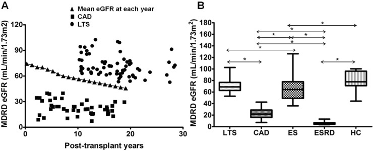
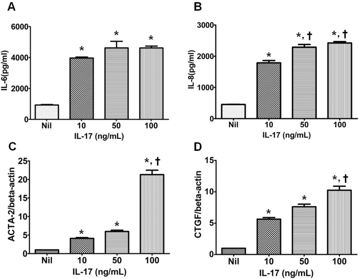
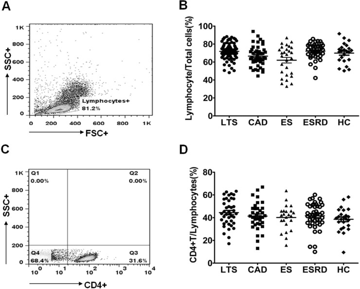
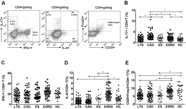
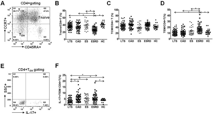
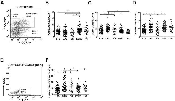
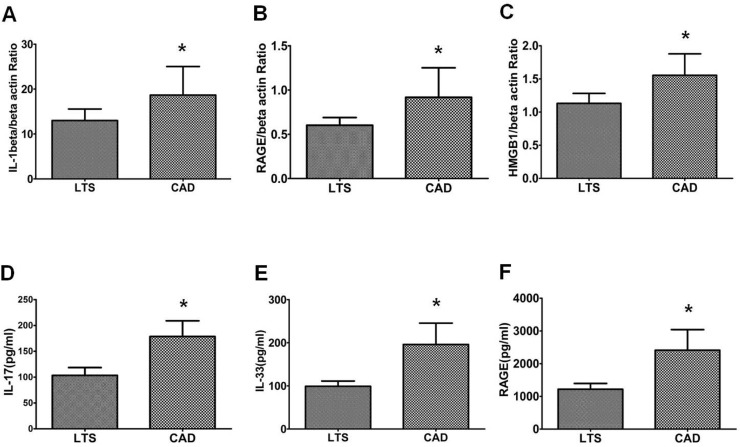
This study was performed to determine the association of Th17 cell phenotype with chronic allograft dysfunction in kidney transplant recipients (KTRs). We compared the expression of Th17 cell phenotype in KTRs with chronic allograft dysfunction group (CAD, n = 52) with four control groups (long-term stable KTRs (LTS, n = 67), early stable KTRs (ES, n = 28), end stage renal disease (ESRD, n = 45), and healthy control (HC, n = 26). We also performed in vitro study using human proximal renal tubular epithelial cell line (HPRTEpiC) to evaluate the effect of IL-17 on human renal tubular epithelial cells. The CAD group showed increased percentage of Th17 cells out of CD4+ T cells and also increased proportion of IL-17 producing cells out of effector memory T cells or out of CCR4+CCR6+/CD4+ T cells compared to the LTS group and other control groups. Also, the serum level of IL-17, IL-33, and RAGE, and the expression of IL-1beta, RAGE, and HMGB1 mRNA showed an increase in the CAD group compared to the LTS group. In vitro study revealed that IL-17 increased production of IL-6 and IL-8 and up-regulated profibrotic gene expression such as ACTA-2 and CTGF in HPRTEpiC in a dose-dependent manner, which suggests that IL-17 has a role in the development of renal tubular cell injury. The results of our study may suggest that increase of Th17 cell phenotype could be a marker for the chronic allograft injury; hence there is a need to develop diagnostic and therapeutic tools targeting the Th17 cells pathway.
本研究旨在确定肾移植受者(KTRs)中Th17细胞表型与慢性移植肾功能障碍之间的关联。我们比较了慢性移植肾功能障碍组(CAD,n = 52)与四个对照组(长期稳定的KTRs(LTS,n = 67)、早期稳定的KTRs(ES,n = 28)、终末期肾病(ESRD,n = 45)和健康对照(HC,n = 26))中KTRs的Th17细胞表型表达。我们还使用人近端肾小管上皮细胞系(HPRTEpiC)进行了体外研究,以评估IL-17对人肾小管上皮细胞的影响。与LTS组和其他对照组相比,CAD组CD4 + T细胞中Th17细胞的百分比增加,效应记忆T细胞或CCR4 + CCR6 + / CD4 + T细胞中产生IL-17的细胞比例也增加。此外,与LTS组相比,CAD组血清IL-17、IL-33和RAGE水平以及IL-1β、RAGE和HMGB1 mRNA表达增加。体外研究表明,IL-17以剂量依赖性方式增加HPRTEpiC中IL-6和IL-8的产生,并上调ACTA-2和CTGF等促纤维化基因的表达,这表明IL-17在肾小管细胞损伤的发生中起作用。我们的研究结果可能表明,Th17细胞表型的增加可能是慢性移植肾损伤的一个标志物;因此,需要开发针对Th17细胞途径的诊断和治疗工具。