• 文献检索
  • 文档翻译
  • 深度研究
  • 学术资讯
  • Suppr Zotero 插件Zotero 插件
  • 邀请有礼
  • 套餐&价格
  • 历史记录
应用&插件
Suppr Zotero 插件Zotero 插件浏览器插件Mac 客户端Windows 客户端微信小程序
定价
高级版会员购买积分包购买API积分包
服务
文献检索文档翻译深度研究API 文档MCP 服务
关于我们
关于 Suppr公司介绍联系我们用户协议隐私条款
关注我们

Suppr 超能文献

核心技术专利:CN118964589B侵权必究
粤ICP备2023148730 号-1Suppr @ 2026

文献检索

告别复杂PubMed语法,用中文像聊天一样搜索,搜遍4000万医学文献。AI智能推荐,让科研检索更轻松。

立即免费搜索

文件翻译

保留排版,准确专业,支持PDF/Word/PPT等文件格式,支持 12+语言互译。

免费翻译文档

深度研究

AI帮你快速写综述,25分钟生成高质量综述,智能提取关键信息,辅助科研写作。

立即免费体验

健康志愿者中心脏扩散张量成像参数与人体测量学之间的关系。

Relationship between cardiac diffusion tensor imaging parameters and anthropometrics in healthy volunteers.

作者信息

McGill L A, Ferreira P F, Scott A D, Nielles-Vallespin S, Giannakidis A, Kilner P J, Gatehouse P D, de Silva R, Firmin D N, Pennell D J

机构信息

NIHR Cardiovascular Biomedical Research Unit, Royal Brompton Hospital, Sydney Street, London, SW3 6NP, UK.

National Heart and Lung Institute, Imperial College, London, UK.

出版信息

J Cardiovasc Magn Reson. 2016 Jan 6;18:2. doi: 10.1186/s12968-015-0215-0.

DOI:10.1186/s12968-015-0215-0
PMID:26738482
原文链接:https://pmc.ncbi.nlm.nih.gov/articles/PMC4704390/
Abstract

BACKGROUND

In vivo cardiac diffusion tensor imaging (cDTI) is uniquely capable of interrogating laminar myocardial dynamics non-invasively. A comprehensive dataset of quantative parameters and comparison with subject anthropometrics is required.

METHODS

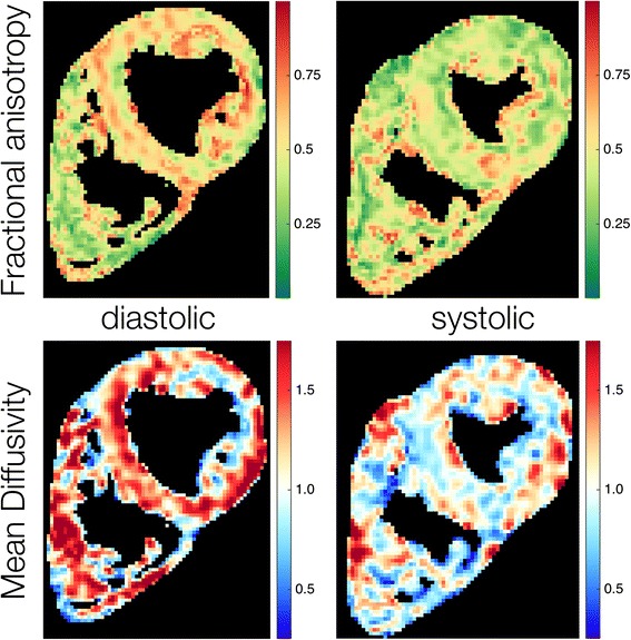
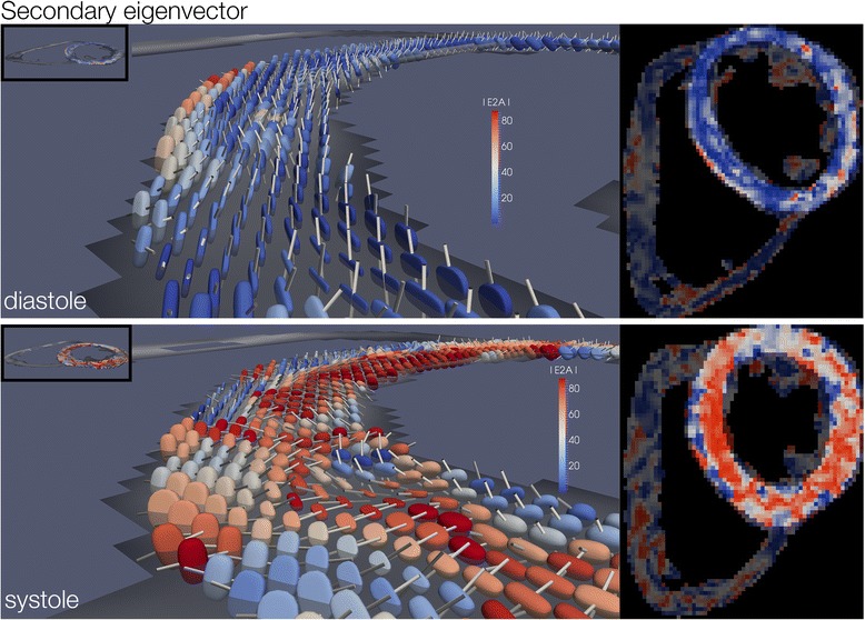
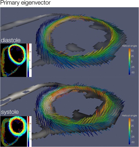
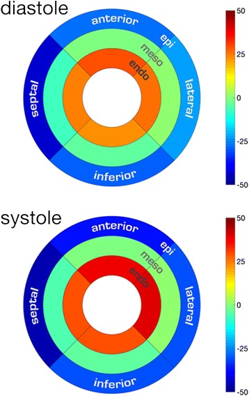
cDTI was performed at 3T with a diffusion weighted STEAM sequence. Data was acquired from the mid left ventricle in 43 subjects during the systolic and diastolic pauses. Global and regional values were determined for fractional anisotropy (FA), mean diffusivity (MD), helix angle gradient (HAg, degrees/%depth) and the secondary eigenvector angulation (E2A). Regression analysis was performed between global values and subject anthropometrics.

RESULTS

All cDTI parameters displayed regional heterogeneity. The RR interval had a significant, but clinically small effect on systolic values for FA, HAg and E2A. Male sex and increasing left ventricular end diastolic volume were associated with increased systolic HAg. Diastolic HAg and systolic E2A were both directly related to left ventricular mass and body surface area. There was an inverse relationship between E2A mobility and both age and ejection fraction.

CONCLUSIONS

Future interpretations of quantitative cDTI data should take into account anthropometric variations observed with patient age, body surface area and left ventricular measurements. Further work determining the impact of technical factors such as strain and SNR is required.

摘要

背景

体内心脏扩散张量成像(cDTI)能够独特地以非侵入性方式研究心肌层动力学。需要一个包含定量参数的综合数据集,并与受试者人体测量学进行比较。

方法

使用扩散加权STEAM序列在3T下进行cDTI。在43名受试者的收缩期和舒张期停顿期间,从左心室中部采集数据。确定了分数各向异性(FA)、平均扩散率(MD)、螺旋角梯度(HAg,度/%深度)和次要特征向量角度(E2A)的整体和区域值。对整体值与受试者人体测量学进行了回归分析。

结果

所有cDTI参数均显示出区域异质性。RR间期对FA、HAg和E2A的收缩期值有显著但临床上较小的影响。男性性别和左心室舒张末期容积增加与收缩期HAg增加相关。舒张期HAg和收缩期E2A均与左心室质量和体表面积直接相关。E2A移动性与年龄和射血分数均呈负相关。

结论

未来对定量cDTI数据的解释应考虑到随患者年龄、体表面积和左心室测量值观察到的人体测量学变化。需要进一步开展工作来确定应变和信噪比等技术因素的影响。

https://cdn.ncbi.nlm.nih.gov/pmc/blobs/875f/4704390/644894cb0551/12968_2015_215_Fig4_HTML.jpg
https://cdn.ncbi.nlm.nih.gov/pmc/blobs/875f/4704390/16230ae2a31f/12968_2015_215_Fig1_HTML.jpg
https://cdn.ncbi.nlm.nih.gov/pmc/blobs/875f/4704390/492cb9500d57/12968_2015_215_Fig2_HTML.jpg
https://cdn.ncbi.nlm.nih.gov/pmc/blobs/875f/4704390/cd8084d03957/12968_2015_215_Fig3_HTML.jpg
https://cdn.ncbi.nlm.nih.gov/pmc/blobs/875f/4704390/644894cb0551/12968_2015_215_Fig4_HTML.jpg

相似文献

1
Relationship between cardiac diffusion tensor imaging parameters and anthropometrics in healthy volunteers.健康志愿者中心脏扩散张量成像参数与人体测量学之间的关系。
J Cardiovasc Magn Reson. 2016 Jan 6;18:2. doi: 10.1186/s12968-015-0215-0.
2
An in-vivo comparison of stimulated-echo and motion compensated spin-echo sequences for 3 T diffusion tensor cardiovascular magnetic resonance at multiple cardiac phases.在 3T 扩散张量心血管磁共振的多个心动周期中,使用激发回波和运动补偿自旋回波序列的活体比较。
J Cardiovasc Magn Reson. 2018 Jan 3;20(1):1. doi: 10.1186/s12968-017-0425-8.
3
In vivo cardiovascular magnetic resonance diffusion tensor imaging shows evidence of abnormal myocardial laminar orientations and mobility in hypertrophic cardiomyopathy.体内心血管磁共振扩散张量成像显示肥厚型心肌病中心肌层状取向和运动异常的证据。
J Cardiovasc Magn Reson. 2014 Nov 12;16(1):87. doi: 10.1186/s12968-014-0087-8.
4
Probing cardiomyocyte mobility with multi-phase cardiac diffusion tensor MRI.应用多期心脏扩散张量 MRI 探测心肌细胞的可移动性。
PLoS One. 2020 Nov 12;15(11):e0241996. doi: 10.1371/journal.pone.0241996. eCollection 2020.
5
Acute Microstructural Changes after ST-Segment Elevation Myocardial Infarction Assessed with Diffusion Tensor Imaging.急性 ST 段抬高型心肌梗死患者的磁共振弥散张量成像表现与急性微观结构变化的相关性研究。
Radiology. 2021 Apr;299(1):86-96. doi: 10.1148/radiol.2021203208. Epub 2021 Feb 9.
6
Diffusion tensor cardiovascular magnetic resonance with a spiral trajectory: An in vivo comparison of echo planar and spiral stimulated echo sequences.基于螺旋轨迹的扩散张量心血管磁共振:在体比较回波平面和螺旋激发回波序列。
Magn Reson Med. 2018 Aug;80(2):648-654. doi: 10.1002/mrm.27051. Epub 2017 Dec 19.
7
The relationship between myocardial microstructure and strain in chronic infarction using cardiovascular magnetic resonance diffusion tensor imaging and feature tracking.应用心血管磁共振弥散张量成像和特征追踪技术研究慢性梗死心肌微结构与应变的关系。
J Cardiovasc Magn Reson. 2022 Nov 24;24(1):66. doi: 10.1186/s12968-022-00892-y.
8
Heterogeneity of Fractional Anisotropy and Mean Diffusivity Measurements by In Vivo Diffusion Tensor Imaging in Normal Human Hearts.正常人体心脏活体扩散张量成像中分数各向异性和平均扩散率测量的异质性
PLoS One. 2015 Jul 15;10(7):e0132360. doi: 10.1371/journal.pone.0132360. eCollection 2015.
9
Reproducibility of in-vivo diffusion tensor cardiovascular magnetic resonance in hypertrophic cardiomyopathy.肥厚型心肌病患者活体扩散张量心血管磁共振的可重复性。
J Cardiovasc Magn Reson. 2012 Dec 24;14(1):86. doi: 10.1186/1532-429X-14-86.
10
Studying Dynamic Myofiber Aggregate Reorientation in Dilated Cardiomyopathy Using In Vivo Magnetic Resonance Diffusion Tensor Imaging.利用活体磁共振扩散张量成像研究扩张型心肌病中动态肌纤维聚集体重定向
Circ Cardiovasc Imaging. 2016 Oct;9(10):e005018. doi: 10.1161/CIRCIMAGING.116.005018.

引用本文的文献

1
Cardiac diffusion-weighted and tensor imaging: A consensus statement from the special interest group of the Society for Cardiovascular Magnetic Resonance.心脏扩散加权成像和张量成像:心血管磁共振学会特别兴趣小组的共识声明
J Cardiovasc Magn Reson. 2024 Oct 22;27(1):101109. doi: 10.1016/j.jocmr.2024.101109.
2
Development of a cardiovascular magnetic resonance-compatible large animal isolated heart model for direct comparison of beating and arrested hearts.开发一种心血管磁共振兼容的大动物离体心脏模型,用于直接比较跳动心脏和停跳心脏。
NMR Biomed. 2022 Jul;35(7):e4692. doi: 10.1002/nbm.4692. Epub 2022 Feb 12.
3
3D-Encoded DENSE MRI with Zonal Excitation for Quantifying Biventricular Myocardial Strain During a Breath-Hold.

本文引用的文献

1
Heterogeneity of Fractional Anisotropy and Mean Diffusivity Measurements by In Vivo Diffusion Tensor Imaging in Normal Human Hearts.正常人体心脏活体扩散张量成像中分数各向异性和平均扩散率测量的异质性
PLoS One. 2015 Jul 15;10(7):e0132360. doi: 10.1371/journal.pone.0132360. eCollection 2015.
2
Age- and gender-related normal left ventricular deformation assessed by cardiovascular magnetic resonance feature tracking.通过心血管磁共振特征追踪评估与年龄和性别相关的正常左心室变形
J Cardiovasc Magn Reson. 2015 Mar 10;17(1):25. doi: 10.1186/s12968-015-0123-3.
3
Probing dynamic myocardial microstructure with cardiac magnetic resonance diffusion tensor imaging.
基于区域激励的三维编码 DENSE MRI 技术在屏气状态下定量评估双心室心肌应变
Cardiovasc Eng Technol. 2021 Dec;12(6):589-597. doi: 10.1007/s13239-021-00561-8. Epub 2021 Jul 9.
4
Cardiovascular magnetic resonance imaging: emerging techniques and applications.心血管磁共振成像:新兴技术与应用。
Heart. 2021 May;107(9):697-704. doi: 10.1136/heartjnl-2019-315669. Epub 2021 Jan 5.
5
Cardiac Magnetic Resonance Quantification of Structure-Function Relationships in Heart Failure.心力衰竭中心脏磁共振结构-功能关系的定量评估。
Heart Fail Clin. 2021 Jan;17(1):9-24. doi: 10.1016/j.hfc.2020.08.001. Epub 2020 Oct 28.
6
Diffusion tensor cardiovascular magnetic resonance in hypertrophic cardiomyopathy: a comparison of motion-compensated spin echo and stimulated echo techniques.肥厚型心肌病的扩散张量心血管磁共振:运动补偿自旋回波与激发回波技术的比较。
MAGMA. 2020 Jun;33(3):331-342. doi: 10.1007/s10334-019-00799-3. Epub 2019 Nov 22.
7
Journal of Cardiovascular Magnetic Resonance 2017.《心血管磁共振杂志》 2017 年刊
J Cardiovasc Magn Reson. 2018 Dec 28;20(1):89. doi: 10.1186/s12968-018-0518-z.
8
Postmortem cardiac magnetic resonance in sudden cardiac death.猝死后的心脏磁共振成像。
Heart Fail Rev. 2018 Sep;23(5):651-665. doi: 10.1007/s10741-018-9705-0.
9
Quantifying precision in cardiac diffusion tensor imaging with second-order motion-compensated convex optimized diffusion encoding.利用二阶运动补偿凸优化扩散编码定量评估心脏扩散张量成像中的精度。
Magn Reson Med. 2018 Sep;80(3):1074-1087. doi: 10.1002/mrm.27107. Epub 2018 Feb 9.
10
An in-vivo comparison of stimulated-echo and motion compensated spin-echo sequences for 3 T diffusion tensor cardiovascular magnetic resonance at multiple cardiac phases.在 3T 扩散张量心血管磁共振的多个心动周期中,使用激发回波和运动补偿自旋回波序列的活体比较。
J Cardiovasc Magn Reson. 2018 Jan 3;20(1):1. doi: 10.1186/s12968-017-0425-8.
利用心脏磁共振扩散张量成像探究动态心肌微观结构
J Cardiovasc Magn Reson. 2014 Nov 12;16(1):89. doi: 10.1186/s12968-014-0089-6.
4
In vivo cardiovascular magnetic resonance diffusion tensor imaging shows evidence of abnormal myocardial laminar orientations and mobility in hypertrophic cardiomyopathy.体内心血管磁共振扩散张量成像显示肥厚型心肌病中心肌层状取向和运动异常的证据。
J Cardiovasc Magn Reson. 2014 Nov 12;16(1):87. doi: 10.1186/s12968-014-0087-8.
5
In vivo contrast free chronic myocardial infarction characterization using diffusion-weighted cardiovascular magnetic resonance.使用扩散加权心血管磁共振对体内无对比剂的慢性心肌梗死进行特征描述。
J Cardiovasc Magn Reson. 2014 Sep 17;16(1):68. doi: 10.1186/s12968-014-0068-y.
6
Characterization of diffuse fibrosis in the failing human heart via diffusion tensor imaging and quantitative histological validation.通过扩散张量成像和定量组织学验证对衰竭人类心脏中的弥漫性纤维化进行表征。
NMR Biomed. 2014 Nov;27(11):1378-86. doi: 10.1002/nbm.3200. Epub 2014 Sep 9.
7
Dual-phase cardiac diffusion tensor imaging with strain correction.具有应变校正的双相心脏扩散张量成像
PLoS One. 2014 Sep 5;9(9):e107159. doi: 10.1371/journal.pone.0107159. eCollection 2014.
8
Intercentre reproducibility of cardiac apparent diffusion coefficient and fractional anisotropy in healthy volunteers.健康志愿者心脏表观扩散系数和各向异性分数的中心间可重复性。
J Cardiovasc Magn Reson. 2014 May 6;16(1):31. doi: 10.1186/1532-429X-16-31.
9
Age- and sex-based reference limits and clinical correlates of myocardial strain and synchrony: the Framingham Heart Study.基于年龄和性别的心肌应变和同步的参考值及临床相关性:弗雷明汉心脏研究。
Circ Cardiovasc Imaging. 2013 Sep;6(5):692-9. doi: 10.1161/CIRCIMAGING.112.000627. Epub 2013 Aug 5.
10
Pushing the limits of in vivo diffusion MRI for the Human Connectome Project.为人类连接组计划推进活体扩散 MRI 的极限
Neuroimage. 2013 Oct 15;80:220-33. doi: 10.1016/j.neuroimage.2013.05.078. Epub 2013 May 24.