Lin Che-Yu, Tsai Chieh-Chih, Kau Hui-Chuan, Yu Wei-Kuang, Kao Shu-Ching, Liu Catherine Jui-Ling
Department of Ophthalmology, Taipei Veterans General Hospital, No. 201, Sec.2, Shih-Pai Road, Taipei, 11217, Taiwan, R.O.C.
Department of Ophthalmology, School of Medicine, National Yang-Ming University, Taipei, Taiwan.
BMC Ophthalmol. 2016 Jan 11;16:8. doi: 10.1186/s12886-016-0185-5.
Angiomyolipoma is a benign mesenchymal tumor composed of variable amounts of smooth muscle, adipose tissue and thick-walled blood vessels, and usually named PEComas (perivascular epithelioid cell tumors). PEComas share overlapping histopathological features with epithelioid cells along a perivascular distribution and characteristic immunohistochemistry with coexpression of myoid and melanocytic markers (HMB-45 /or Melan-A). We report the first case of primary orbital angiomyolipoma with negative melanocytic marker.
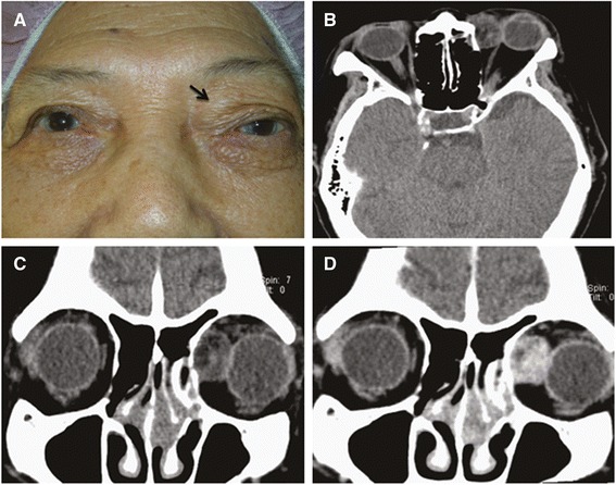
An 80-year-old Asian woman had a 2-year history of progressive swelling in the left upper eyelid. External examination revealed 3 cm of relative proptosis of the left eye and a palpable mass in the left superonasal orbit. Computed tomographic scan demonstrated a circumscribed, heterogeneous orbital mass. Excision biopsy was done and the histological finding demonstrated the orbital mass was composed of mature adipocytes, intermingled with spindle or oval-shaped cells, and accompanied by thick-walled blood vessels. Immunohistochemically, tumor cells were positive for CD34 and HHF-35, but negative for cytokeratin, HMB-45 and Melan-A. The diagnosis of angiomyolipoma was made. No recurrence was noted at 2-year follow-up.
In our case, the HMB-45 negativity may be explained by the rarity of the epithelioid cells, and the HMB-45 positivity is often weaker or absent in spindle cells. Angiomyolipoma, although rare, should be added to the differential diagnosis of space-occupying orbital lesion.
血管平滑肌脂肪瘤是一种良性间叶组织肿瘤,由数量不等的平滑肌、脂肪组织和厚壁血管组成,通常被称为PEComas(血管周上皮样细胞瘤)。PEComas具有沿血管分布的上皮样细胞重叠的组织病理学特征以及肌样和黑素细胞标志物(HMB-45 /或Melan-A)共表达的特征性免疫组化表现。我们报告首例黑素细胞标志物阴性的原发性眼眶血管平滑肌脂肪瘤病例。
一名80岁的亚洲女性有左上睑进行性肿胀2年病史。外部检查显示左眼相对眼球突出3cm,左鼻上眶可触及肿块。计算机断层扫描显示一个边界清楚的、不均匀的眼眶肿块。进行了切除活检,组织学检查发现眼眶肿块由成熟脂肪细胞组成,混有梭形或椭圆形细胞,并伴有厚壁血管。免疫组化显示,肿瘤细胞CD34和HHF-35阳性,但细胞角蛋白、HMB-45和Melan-A阴性。诊断为血管平滑肌脂肪瘤。2年随访未发现复发。
在我们的病例中,HMB-45阴性可能是由于上皮样细胞罕见,且梭形细胞中HMB-45阳性通常较弱或缺失。血管平滑肌脂肪瘤虽然罕见,但应列入眼眶占位性病变的鉴别诊断。