Yang Daniel Y-J, Beam Danielle, Pelphrey Kevin A, Abdullahi Sebiha, Jou Roger J
Center for Translational Developmental Neuroscience, Child Study Center, Yale University, New Haven, CT USA.
Mol Autism. 2016 Jan 25;7:11. doi: 10.1186/s13229-016-0076-x. eCollection 2016.
Individuals with autism spectrum disorder (ASD) have been characterized by altered cerebral cortical structures; however, the field has yet to identify consistent markers and prior studies have included mostly adolescents and adults. While there are multiple cortical morphological measures, including cortical thickness, surface area, cortical volume, and cortical gyrification, few single studies have examined all these measures. The current study analyzed all of the four measures and focused on pre-adolescent children with ASD.
We employed the FreeSurfer pipeline to examine surface-based morphometry in 60 high-functioning boys with ASD (mean age = 8.35 years, range = 4-12 years) and 41 gender-, age-, and IQ-matched typically developing (TD) peers (mean age = 8.83 years), while testing for age-by-diagnosis interaction and between-group differences.
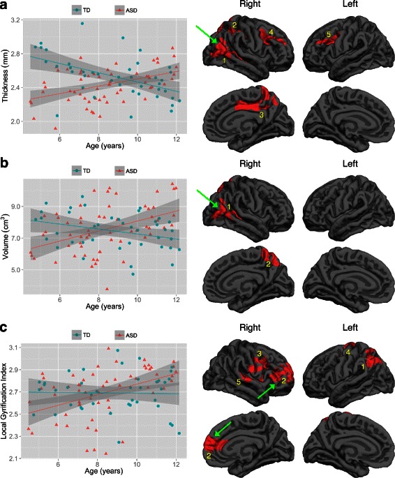
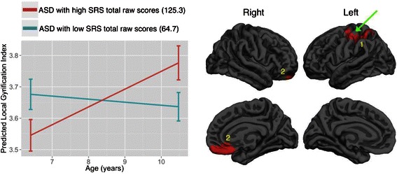
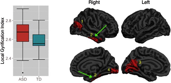
During childhood and in specific regions, ASD participants exhibited a lack of normative age-related cortical thinning and volumetric reduction and an abnormal age-related increase in gyrification. Regarding surface area, ASD and TD exhibited statistically comparable age-related development during childhood. Across childhood, ASD relative to TD participants tended to have higher mean levels of gyrification in specific regions. Within ASD, those with higher Social Responsiveness Scale total raw scores tended to have greater age-related increase in gyrification in specific regions during childhood.
ASD is characterized by cortical neuroanatomical abnormalities that are age-, measure-, statistical model-, and region-dependent. The current study is the first to examine the development of all four cortical measures in one of the largest pre-adolescent samples. Strikingly, Neurosynth-based quantitative reverse inference of the surviving clusters suggests that many of the regions identified above are related to social perception, language, self-referential, and action observation networks-those frequently found to be functionally altered in individuals with ASD. The comprehensive, multilevel analyses across a wide range of cortical measures help fill a knowledge gap and present a complex but rich picture of neuroanatomical developmental differences in children with ASD.
自闭症谱系障碍(ASD)患者具有大脑皮质结构改变的特征;然而,该领域尚未确定一致的标志物,并且先前的研究主要纳入的是青少年和成年人。虽然有多种皮质形态学测量方法,包括皮质厚度、表面积、皮质体积和皮质回旋,但很少有单一研究对所有这些测量方法进行考察。本研究分析了所有这四种测量方法,并聚焦于患有ASD的青春期前儿童。
我们采用FreeSurfer流程来检查60名高功能ASD男孩(平均年龄 = 8.35岁,范围 = 4 - 12岁)和41名性别、年龄和智商匹配的发育正常(TD)同龄人(平均年龄 = 8.83岁)的基于表面的形态学,同时测试年龄与诊断的交互作用和组间差异。
在儿童期及特定区域,ASD参与者表现出缺乏与年龄相关的正常皮质变薄和体积减少,以及与年龄相关的回旋异常增加。关于表面积,ASD和TD在儿童期表现出在统计学上可比的与年龄相关的发育。在整个儿童期,与TD参与者相比,ASD参与者在特定区域往往具有更高的平均回旋水平。在ASD患者中,社交反应量表总原始得分较高的个体在儿童期特定区域往往具有更大的与年龄相关的回旋增加。
ASD的特征是皮质神经解剖学异常,这些异常与年龄、测量方法、统计模型和区域有关。本研究是首次在最大的青春期前样本之一中考察所有四种皮质测量方法的发育情况。引人注目的是,基于Neurosynth对存活簇的定量反向推断表明,上述许多区域与社会感知、语言、自我参照和动作观察网络有关,这些网络在ASD个体中经常被发现功能改变。对广泛的皮质测量方法进行全面、多层次分析有助于填补知识空白,并呈现出ASD儿童神经解剖学发育差异的复杂但丰富的图景。