Vetreno Ryan P, Yaxley Richard, Paniagua Beatriz, Johnson G Allan, Crews Fulton T
Bowles Center for Alcohol Studies, Department of Psychiatry, University of North Carolina at Chapel Hill, Chapel Hill, NC, USA.
Duke Center for In Vivo Microscopy, Duke University Medical Center, Durham, NC, USA.
Addict Biol. 2017 May;22(3):712-723. doi: 10.1111/adb.12364. Epub 2016 Feb 1.
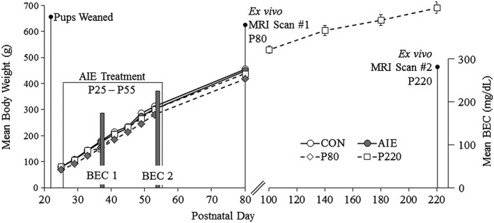
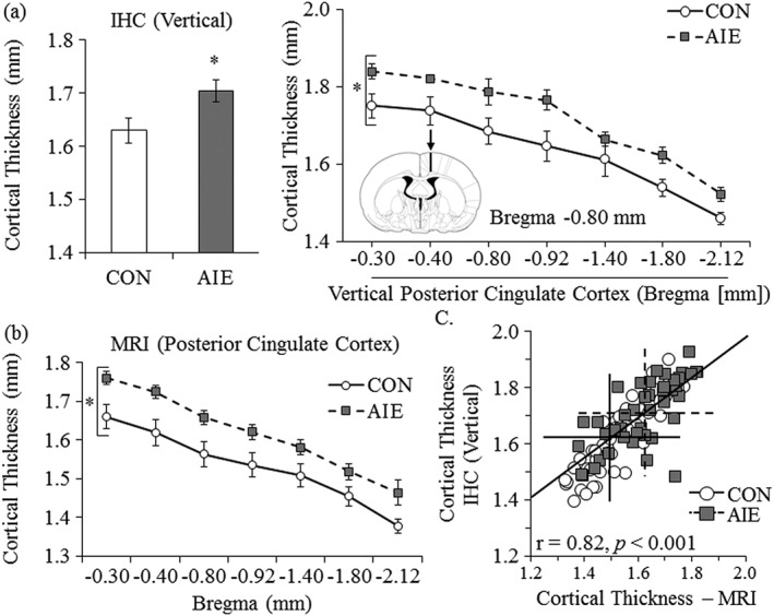
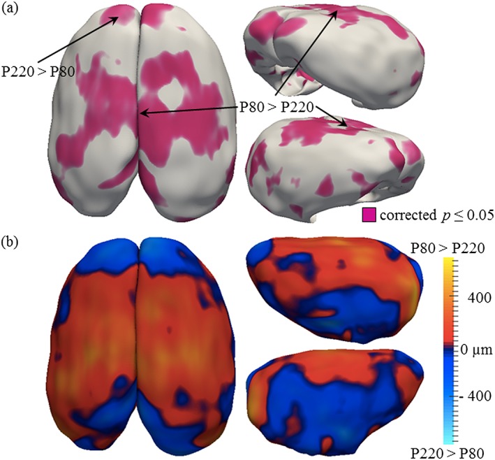
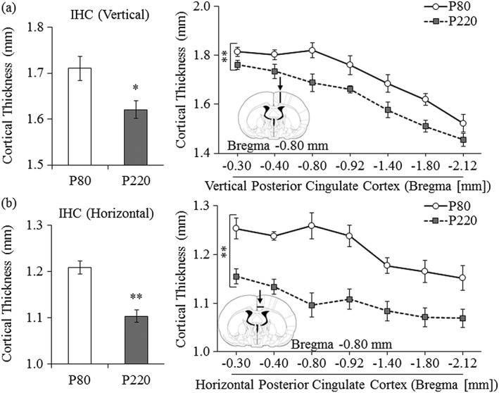
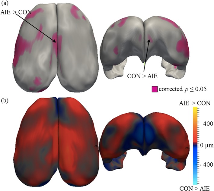
Human studies have established that adolescence is a period of brain maturation that parallels the development of adult behaviors. However, little is known regarding cortical development in the adult rat brain. We used magnetic resonance imaging (MRI) and histology to assess the impact of age on adult Wistar rat cortical thickness on postnatal day (P)80 and P220 as well as the effect of adolescent binge ethanol exposure on adult (P80) cortical thickness. MRI revealed changes in cortical thickness between P80 and P220 that differ across cortical region. The adult P220 rat prefrontal cortex increased in thickness whereas cortical thinning occurred in both the cingulate and parietal cortices relative to young adult P80 rats. Histological analysis confirmed the age-related cortical thinning. In the second series of experiments, an animal model of adolescent intermittent ethanol (AIE; 5.0 g/kg, intragastrically, 20 percent ethanol w/v, 2 days on/2 days off from P25 to P55) was used to assess the effects of alcohol on cortical thickness in young adult (P80) rats. MRI revealed that AIE resulted in region-specific cortical changes. A small region within the prefrontal cortex was significantly thinner whereas medial cortical regions were significantly thicker in young adult (P80) AIE-treated rats. The observed increase in cortical thickness was confirmed by histology. Thus, the rat cerebral cortex continues to undergo cortical thickness changes into adulthood, and adolescent alcohol exposure alters the young adult cortex that could contribute to brain dysfunction in adulthood.
人体研究已经证实,青春期是大脑成熟的时期,这与成人行为的发展并行。然而,关于成年大鼠大脑皮质发育的情况却知之甚少。我们使用磁共振成像(MRI)和组织学方法,评估年龄对出生后第80天(P80)和第220天(P220)成年Wistar大鼠皮质厚度的影响,以及青少年暴饮乙醇对成年(P80)大鼠皮质厚度的影响。MRI显示,P80和P220之间皮质厚度的变化因皮质区域而异。成年P220大鼠的前额叶皮质厚度增加,而与年轻成年P80大鼠相比,扣带回和顶叶皮质均出现皮质变薄。组织学分析证实了与年龄相关的皮质变薄。在第二系列实验中,使用青少年间歇性乙醇(AIE;5.0克/千克,灌胃,20%乙醇重量/体积,从P25至P55,2天给药/2天停药)动物模型,评估酒精对年轻成年(P80)大鼠皮质厚度的影响。MRI显示,AIE导致了区域特异性的皮质变化。在年轻成年(P80)接受AIE治疗的大鼠中,前额叶皮质内的一个小区域明显变薄,而内侧皮质区域明显增厚。组织学证实了观察到的皮质厚度增加。因此,大鼠大脑皮质在成年期仍会持续发生皮质厚度变化,青少年酒精暴露会改变年轻成年大鼠的皮质,这可能导致成年期脑功能障碍。