Szigeti Krisztián, Szabó Tibor, Korom Csaba, Czibak Ilona, Horváth Ildikó, Veres Dániel S, Gyöngyi Zoltán, Karlinger Kinga, Bergmann Ralf, Pócsik Márta, Budán Ferenc, Máthé Domokos
Department of Biophysics and Radiation Biology, Semmelweis University, Tűzoltó utca 37-47, Budapest, H-1094, Hungary.
CROmed Translational Research Centers Ltd., Baross utca 91-95, Budapest, H-1047, Hungary.
BMC Med Imaging. 2016 Feb 11;16:14. doi: 10.1186/s12880-016-0118-z.
Lung diseases (resulting from air pollution) require a widely accessible method for risk estimation and early diagnosis to ensure proper and responsive treatment. Radiomics-based fractal dimension analysis of X-ray computed tomography attenuation patterns in chest voxels of mice exposed to different air polluting agents was performed to model early stages of disease and establish differential diagnosis.
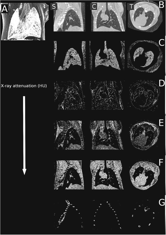
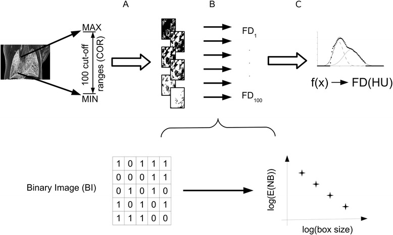
To model different types of air pollution, BALBc/ByJ mouse groups were exposed to cigarette smoke combined with ozone, sulphur dioxide gas and a control group was established. Two weeks after exposure, the frequency distributions of image voxel attenuation data were evaluated. Specific cut-off ranges were defined to group voxels by attenuation. Cut-off ranges were binarized and their spatial pattern was associated with calculated fractal dimension, then abstracted by the fractal dimension -- cut-off range mathematical function. Nonparametric Kruskal-Wallis (KW) and Mann-Whitney post hoc (MWph) tests were used.
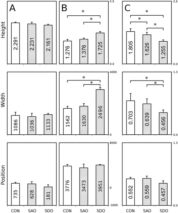
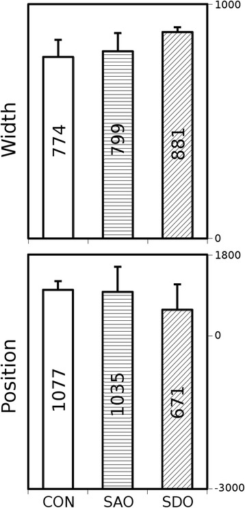
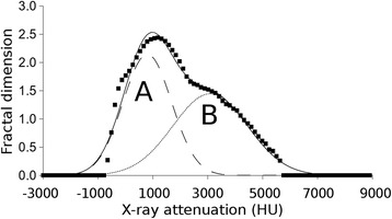
Each cut-off range versus fractal dimension function plot was found to contain two distinctive Gaussian curves. The ratios of the Gaussian curve parameters are considerably significant and are statistically distinguishable within the three exposure groups.
A new radiomics evaluation method was established based on analysis of the fractal dimension of chest X-ray computed tomography data segments. The specific attenuation patterns calculated utilizing our method may diagnose and monitor certain lung diseases, such as chronic obstructive pulmonary disease (COPD), asthma, tuberculosis or lung carcinomas.
肺部疾病(由空气污染导致)需要一种广泛可用的风险评估和早期诊断方法,以确保进行适当且及时的治疗。对暴露于不同空气污染物的小鼠胸部体素的X射线计算机断层扫描衰减模式进行基于放射组学的分形维数分析,以模拟疾病的早期阶段并建立鉴别诊断。
为了模拟不同类型的空气污染,将BALBc/ByJ小鼠组暴露于香烟烟雾与臭氧的组合中,建立了二氧化硫气体组和一个对照组。暴露两周后,评估图像体素衰减数据的频率分布。通过衰减定义特定的截断范围以对体素进行分组。截断范围进行二值化处理,其空间模式与计算出的分形维数相关联,然后通过分形维数 - 截断范围数学函数进行提取。使用非参数Kruskal-Wallis(KW)和Mann-Whitney事后检验(MWph)。
发现每个截断范围与分形维数函数图包含两条独特的高斯曲线。高斯曲线参数的比率相当显著,并且在三个暴露组内具有统计学差异。
基于对胸部X射线计算机断层扫描数据段分形维数的分析,建立了一种新的放射组学评估方法。利用我们的方法计算出的特定衰减模式可用于诊断和监测某些肺部疾病,如慢性阻塞性肺疾病(COPD)、哮喘、肺结核或肺癌。