Bulut Mehmet Deniz, Alpayci Mahmut, Şenköy Emre, Bora Aydin, Yazmalar Levent, Yavuz Alpaslan, Gülşen İsmail
Department of Radiology, Yuzuncu Yil University, Medical Faculty, Van, Turkey.
Department of Physical Medicine and Rehabilitation, Yuzuncu Yil University, Medical Faculty, Van, Turkey.
Med Sci Monit. 2016 Feb 15;22:495-500. doi: 10.12659/msm.897500.
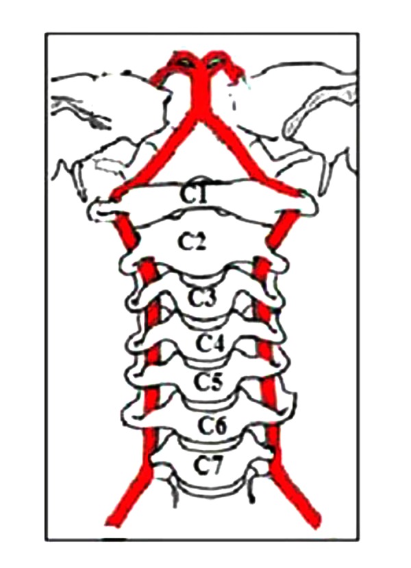
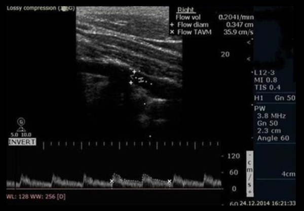
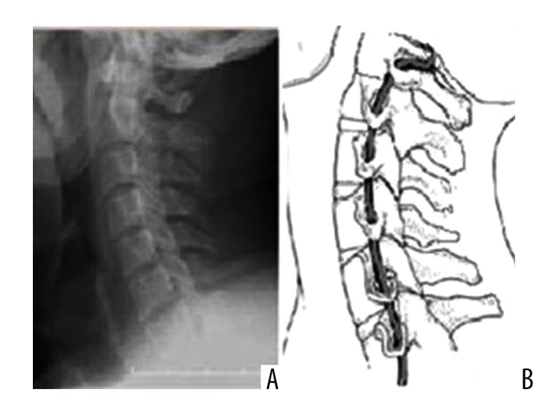
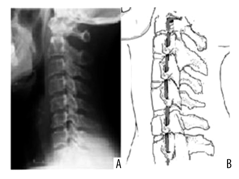
BACKGROUND Because loss of cervical lordosis leads to disrupted biomechanics, the natural lordotic curvature is considered to be an ideal posture for the cervical spine. The vertebral arteries proceed in the transverse foramen of each cervical vertebra. Considering that the vertebral arteries travel in close anatomical relationship to the cervical spine, we speculated that the loss of cervical lordosis may affect vertebral artery hemodynamics. The aim of this study was to compare the vertebral artery values between subjects with and without loss of cervical lordosis. MATERIAL AND METHODS Thirty patients with loss of cervical lordosis and 30 controls matched for age, sex, and body mass index were included in the study. Sixty vertebral arteries in patients with loss of cervical lordosis and 60 in controls without loss of cervical lordosis were evaluated by Doppler ultrasonography. Vertebral artery hemodynamics, including lumen diameter, flow volume, peak systolic velocity, end-diastolic velocity, and resistive index, were measured, and determined values were statistically compared between the patient and the control groups. RESULTS The means of diameter (p=0.003), flow volume (p=0.002), and peak systolic velocity (p=0.014) in patients were significantly lower as compared to controls. However, there was no significant difference between the 2 groups in terms of the end-diastolic velocity (p=0.276) and resistive index (p=0.536) parameters. CONCLUSIONS The present study revealed a significant association between loss of cervical lordosis and decreased vertebral artery hemodynamics, including diameter, flow volume, and peak systolic velocity. Further studies are required to confirm these findings and to investigate their possible clinical implications.
由于颈椎生理前凸消失会导致生物力学紊乱,因此自然的前凸曲度被认为是颈椎的理想姿势。椎动脉走行于各颈椎的横突孔内。考虑到椎动脉与颈椎在解剖学上关系密切,我们推测颈椎生理前凸消失可能会影响椎动脉血流动力学。本研究的目的是比较有和没有颈椎生理前凸消失的受试者之间的椎动脉参数值。
本研究纳入了30例颈椎生理前凸消失的患者以及30例年龄、性别和体重指数相匹配的对照组。采用多普勒超声对30例颈椎生理前凸消失患者的60条椎动脉以及30例无颈椎生理前凸消失对照组的60条椎动脉进行评估。测量椎动脉血流动力学参数,包括管腔直径、血流量、收缩期峰值流速、舒张末期流速和阻力指数,并对患者组和对照组的测定值进行统计学比较。
与对照组相比,患者的管腔直径(p=0.003)、血流量(p=0.002)和收缩期峰值流速(p=0.014)均值显著降低。然而,两组在舒张末期流速(p=0.276)和阻力指数(p=0.536)参数方面无显著差异。
本研究揭示了颈椎生理前凸消失与椎动脉血流动力学降低之间存在显著关联,包括管腔直径、血流量和收缩期峰值流速。需要进一步研究来证实这些发现并探讨其可能的临床意义。