Department of Medical Physiology, College of Medicine, Texas A&M University, Bryan, TX, United States of America.
Gastroenterology, Medicine, Indiana University, Indianapolis, IN, United States of America.
EBioMedicine. 2019 Oct;48:130-142. doi: 10.1016/j.ebiom.2019.09.013. Epub 2019 Sep 12.
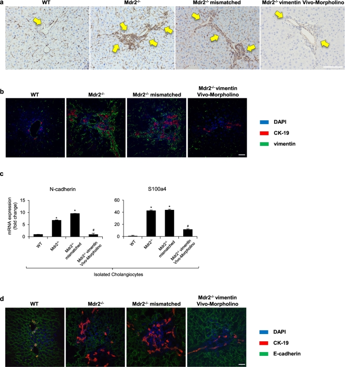
Cholangiocytes are the target cells of cholangiopathies including primary sclerosing cholangitis (PSC). Vimentin is an intermediate filament protein that has been found in various types of mesenchymal cells. The aim of this study is to evaluate the role of vimentin in the progression of biliary damage/liver fibrosis and whether there is a mesenchymal phenotype of cholangiocytes in the Mdr2 model of PSC.
In vivo studies were performed in 12 wk. Mdr2 male mice with or without vimentin Vivo-Morpholino treatment and their corresponding control groups. Liver specimens from human PSC patients, human intrahepatic biliary epithelial cells (HIBEpiC) and human hepatic stellate cell lines (HHSteCs) were used to measure changes in epithelial-to-mesenchymal transition (EMT).
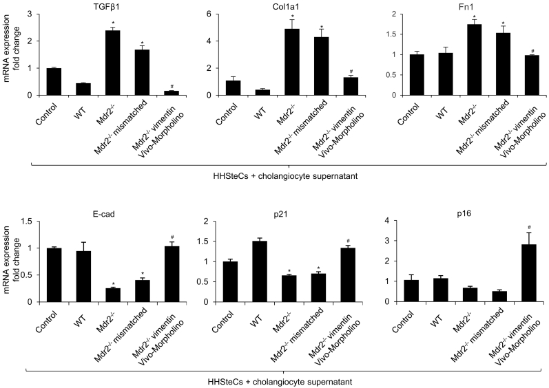
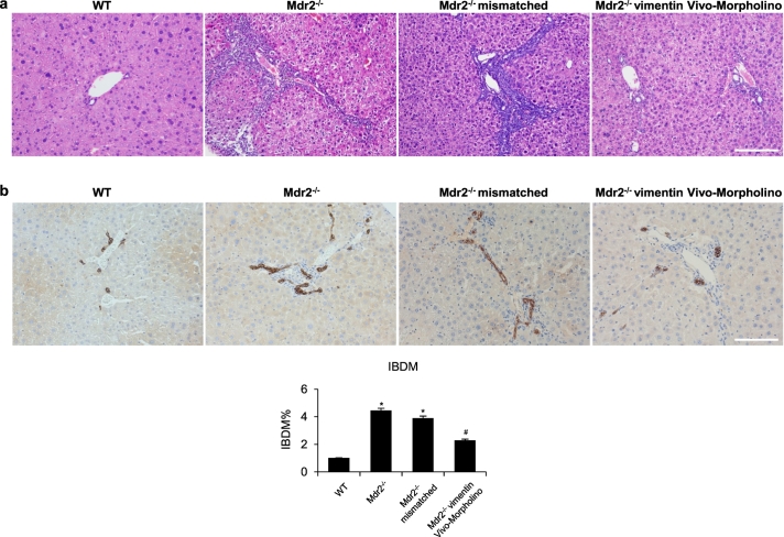
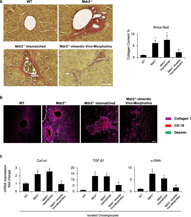
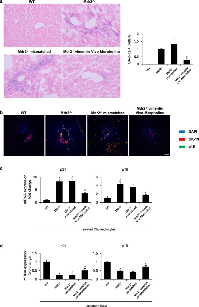
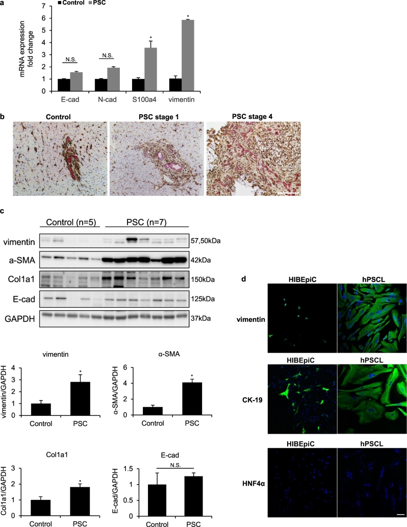
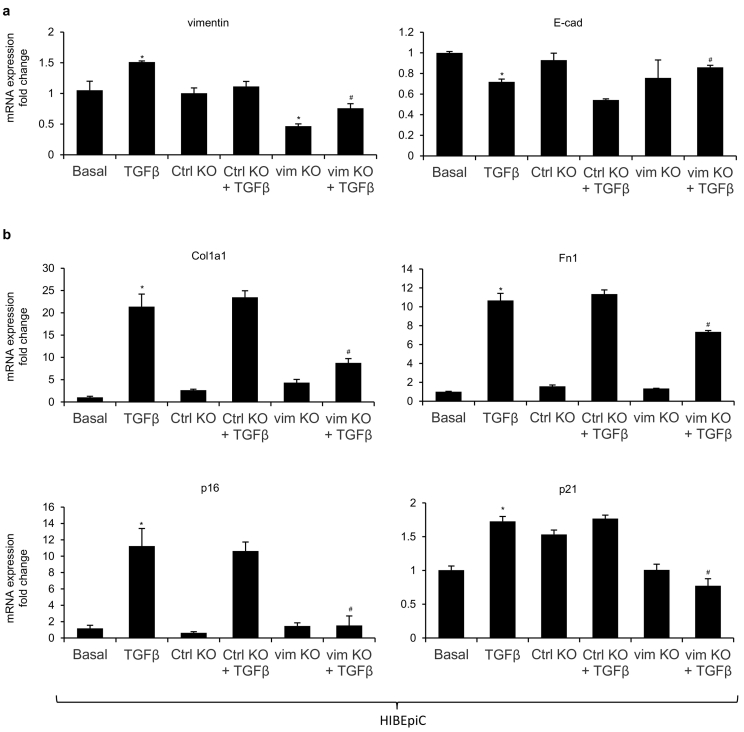
There was increased mesenchymal phenotype of cholangiocytes in Mdr2 mice, which was reduced by treatment of vimentin Vivo-Morpholino. Concomitant with reduced vimentin expression, there was decreased liver damage, ductular reaction, biliary senescence, liver fibrosis and TGF-β1 secretion in Mdr2 mice treated with vimentin Vivo-Morpholino. Human PSC patients and derived cell lines had increased expression of vimentin and other mesenchymal markers compared to healthy controls and HIBEpiC, respectively. In vitro silencing of vimentin in HIBEpiC suppressed TGF-β1-induced EMT and fibrotic reaction. HHSteCs had decreased fibrotic reaction and increased cellular senescence after stimulation with cholangiocyte supernatant with reduced vimentin levels.
Our study demonstrated that knockdown of vimentin reduces mesenchymal phenotype of cholangiocytes, which leads to decreased biliary senescence and liver fibrosis. Inhibition of vimentin may be a key therapeutic target in the treatment of cholangiopathies including PSC. FUND: National Institutes of Health (NIH) awards, VA Merit awards.
胆管细胞是包括原发性硬化性胆管炎(PSC)在内的胆管疾病的靶细胞。波形蛋白是一种中间丝蛋白,已在各种类型的间充质细胞中发现。本研究旨在评估波形蛋白在胆汁损伤/肝纤维化进展中的作用,以及在 Mdr2 模型的 PSC 中是否存在胆管细胞的间充质表型。
在 12 周内进行体内研究。Mdr2 雄性小鼠,有无波形蛋白 Vivo-Morpholino 处理及其相应的对照组。使用来自人 PSC 患者、人肝内胆管上皮细胞(HIBEpiC)和人肝星状细胞系(HHSteCs)的肝组织标本来测量上皮-间质转化(EMT)的变化。
在 Mdr2 小鼠中观察到胆管细胞的间充质表型增加,用波形蛋白 Vivo-Morpholino 处理可减少该表型。与波形蛋白表达减少同时,Mdr2 小鼠用 Vivo-Morpholino 处理后,肝损伤、胆管反应、胆管衰老、肝纤维化和 TGF-β1 分泌减少。与健康对照组和 HIBEpiC 相比,人 PSC 患者和衍生细胞系的波形蛋白和其他间充质标志物表达增加。在 HIBEpiC 中沉默波形蛋白可抑制 TGF-β1 诱导的 EMT 和纤维反应。在刺激用降低了波形蛋白水平的胆管细胞上清液后,HHSteCs 的纤维化反应减少,细胞衰老增加。
我们的研究表明,敲低波形蛋白可减少胆管细胞的间充质表型,从而减少胆汁衰老和肝纤维化。抑制波形蛋白可能是治疗包括 PSC 在内的胆管疾病的关键治疗靶点。
美国国立卫生研究院(NIH)奖、VA 功绩奖。