Okani Chukwudi Onyeaghana, Otene Benjamin, Nyaga Terhemba, Ngbea Joseph, Eke Agaba, Edegbe Felix, Anyiam Daniel
Department of Histopathology, Chukwuemeka Odumegwu Ojukwu University, Awka Campus, Nigeria.
Department of Histopathology, Benue State University Teaching Hospital, Makurdi, Benue State, Nigeria.
Niger Med J. 2015 Nov-Dec;56(6):433-5. doi: 10.4103/0300-1652.171612.
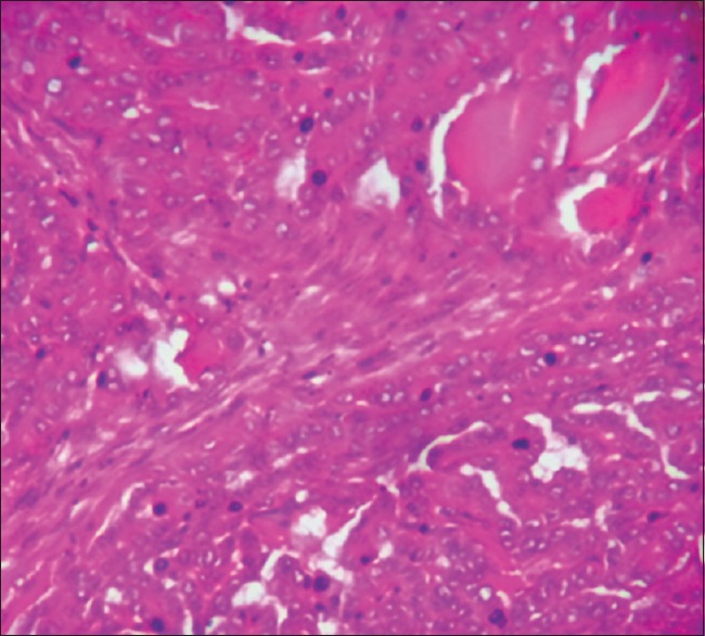
Hashimoto thyroiditis (HT) is an autoimmune disease, known to be the most common cause of hypothyroidism in nonendemic goitrous areas. It is usually characterized by symmetric, painless, and diffused but sometimes localized swelling of the thyroid gland with features of hypothyroidism. Papillary thyroid carcinoma (PTC), on the other hand, is the most common yet less aggressive form of thyroid cancer, especially in iodine-deficient areas. The coexistence of the two diseases is possible but not common. This case study reports a 50-year-old female with a 10-year history of a huge goiter, which was essentially symptom-free until about 3 months prior to presentation when the patient started complaining of neck pain, dysphagia, productive cough, and cold intolerance. Physical examination revealed focal cystic and tender area in the multinodular swelling and associated cervical lymphadenopathy on the left side of the neck. The serum thyroid stimulating hormone was high, sub-normal T3, and the T4 was low. The fine needle aspiration cytology yielded 10 ml of aspirate of pus admixed with altered blood which on microscopy showed a few suspicious follicular epithelial cells with open nuclei admixed with mainly neutrophil polymorphs, siderophages, and foam cells in a hemorrhagic background. The patient had an incision biopsy that showed areas displaying PTC and HT.
桥本甲状腺炎(HT)是一种自身免疫性疾病,是非地方性甲状腺肿地区甲状腺功能减退最常见的病因。其通常表现为甲状腺呈对称性、无痛性、弥漫性肿大,但有时也可呈局限性肿大,并伴有甲状腺功能减退的特征。另一方面,乳头状甲状腺癌(PTC)是最常见但侵袭性较小的甲状腺癌类型,尤其是在碘缺乏地区。这两种疾病可能同时存在,但并不常见。本病例报告了一名50岁女性,有巨大甲状腺肿10年病史,在就诊前约3个月出现颈部疼痛、吞咽困难、咳痰和畏寒等症状之前基本无症状。体格检查发现多结节性肿大处有局灶性囊性和压痛区,左侧颈部伴有颈淋巴结病。血清促甲状腺激素升高,T3低于正常,T4降低。细针穿刺细胞学检查吸出10毫升混有血性液体的脓液,显微镜检查显示有一些可疑的滤泡上皮细胞,细胞核开放,在出血背景中主要混有中性多形核白细胞、含铁血黄素细胞和泡沫细胞。患者进行了切开活检,结果显示存在PTC和HT区域。