Nolan Ryan M, Adie Steven G, Marjanovic Marina, Chaney Eric J, South Fredrick A, Monroy Guillermo L, Shemonski Nathan D, Erickson-Bhatt Sarah J, Shelton Ryan L, Bower Andrew J, Simpson Douglas G, Cradock Kimberly A, Liu Z George, Ray Partha S, Boppart Stephen A
Beckman Institute for Advanced Science and Technology, University of Illinois at Urbana-Champaign (UIUC), 405 N. Mathews Ave., Urbana, IL, 61801, USA.
PhotoniCare, Inc., Champaign, IL, USA.
BMC Cancer. 2016 Feb 23;16:144. doi: 10.1186/s12885-016-2194-4.
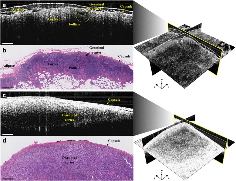
Evaluation of lymph node (LN) status is an important factor for detecting metastasis and thereby staging breast cancer. Currently utilized clinical techniques involve the surgical disruption and resection of lymphatic structure, whether nodes or axillary contents, for histological examination. While reasonably effective at detection of macrometastasis, the majority of the resected lymph nodes are histologically negative. Improvements need to be made to better detect micrometastasis, minimize or eliminate lymphatic disruption complications, and provide immediate and accurate intraoperative feedback for in vivo cancer staging to better guide surgery.
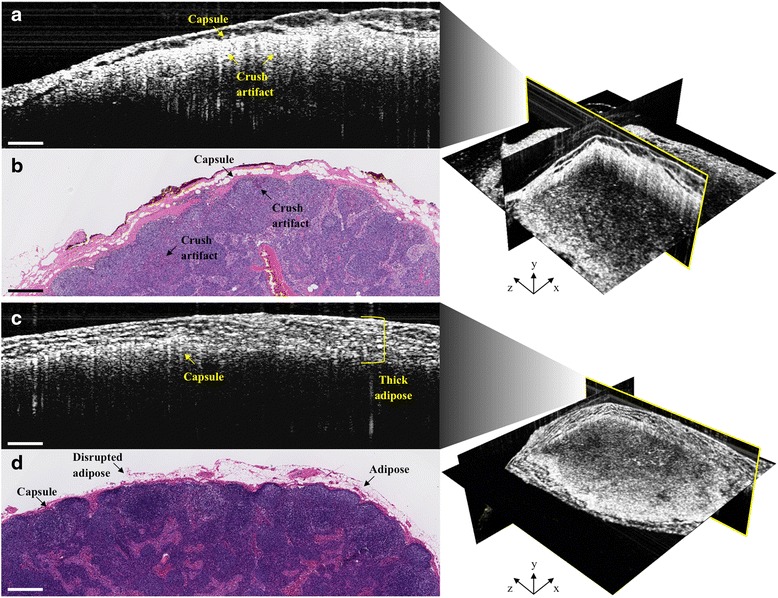
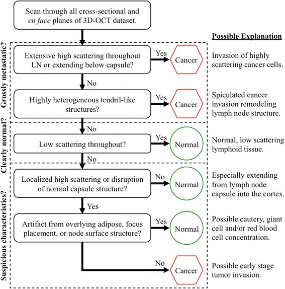
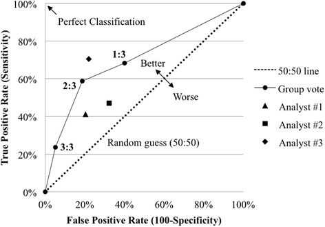
We evaluated the use of optical coherence tomography (OCT), a high-resolution, real-time, label-free imaging modality for the intraoperative assessment of human LNs for metastatic disease in patients with breast cancer. We assessed the sensitivity and specificity of double-blinded trained readers who analyzed intraoperative OCT LN images for presence of metastatic disease, using co-registered post-operative histopathology as the gold standard.
Our results suggest that intraoperative OCT examination of LNs is an appropriate real-time, label-free, non-destructive alternative to frozen-section analysis, potentially offering faster interpretation and results to empower superior intraoperative decision-making.
Intraoperative OCT has strong potential to supplement current post-operative histopathology with real-time in situ assessment of LNs to preserve both non-cancerous nodes and their lymphatic vessels, and thus reduce the associated risks and complications from surgical disruption of lymphoid structures following biopsy.
评估淋巴结(LN)状态是检测转移从而对乳腺癌进行分期的重要因素。目前使用的临床技术包括对淋巴结构(无论是淋巴结还是腋窝内容物)进行手术破坏和切除,以进行组织学检查。虽然在检测大转移灶方面相当有效,但大多数切除的淋巴结在组织学上为阴性。需要改进以更好地检测微转移,最小化或消除淋巴破坏并发症,并为体内癌症分期提供即时准确的术中反馈,以更好地指导手术。
我们评估了光学相干断层扫描(OCT)的应用,这是一种高分辨率、实时、无标记的成像方式,用于在术中评估乳腺癌患者的人淋巴结是否存在转移性疾病。我们使用共同注册的术后组织病理学作为金标准,评估了双盲训练的读者分析术中OCT淋巴结图像中转移性疾病存在情况的敏感性和特异性。
我们的结果表明,术中对淋巴结进行OCT检查是一种合适的实时、无标记、非破坏性的替代冷冻切片分析的方法,可能提供更快的解读和结果,以增强术中决策能力。
术中OCT有很大潜力通过对淋巴结进行实时原位评估来补充当前的术后组织病理学,以保留非癌性淋巴结及其淋巴管,从而降低活检后淋巴结构手术破坏相关的风险和并发症。