Yang Lingxiao, Park Jaena, Marjanovic Marina, Chaney Eric J, Spillman Darold R, Phillips Heidi, Boppart Stephen A
Beckman Institute for Advanced Science and Technology, University of Illinois at Urbana-Champaign, Urbana, IL 61801 USA.
Small Animal Surgery, Veterinary Teaching Hospital, University of Illinois College of Veterinary Medicine, Urbana, IL 61802 USA.
IEEE J Sel Top Quantum Electron. 2021 Jul-Aug;27(4). doi: 10.1109/jstqe.2021.3054578. Epub 2021 Jan 26.
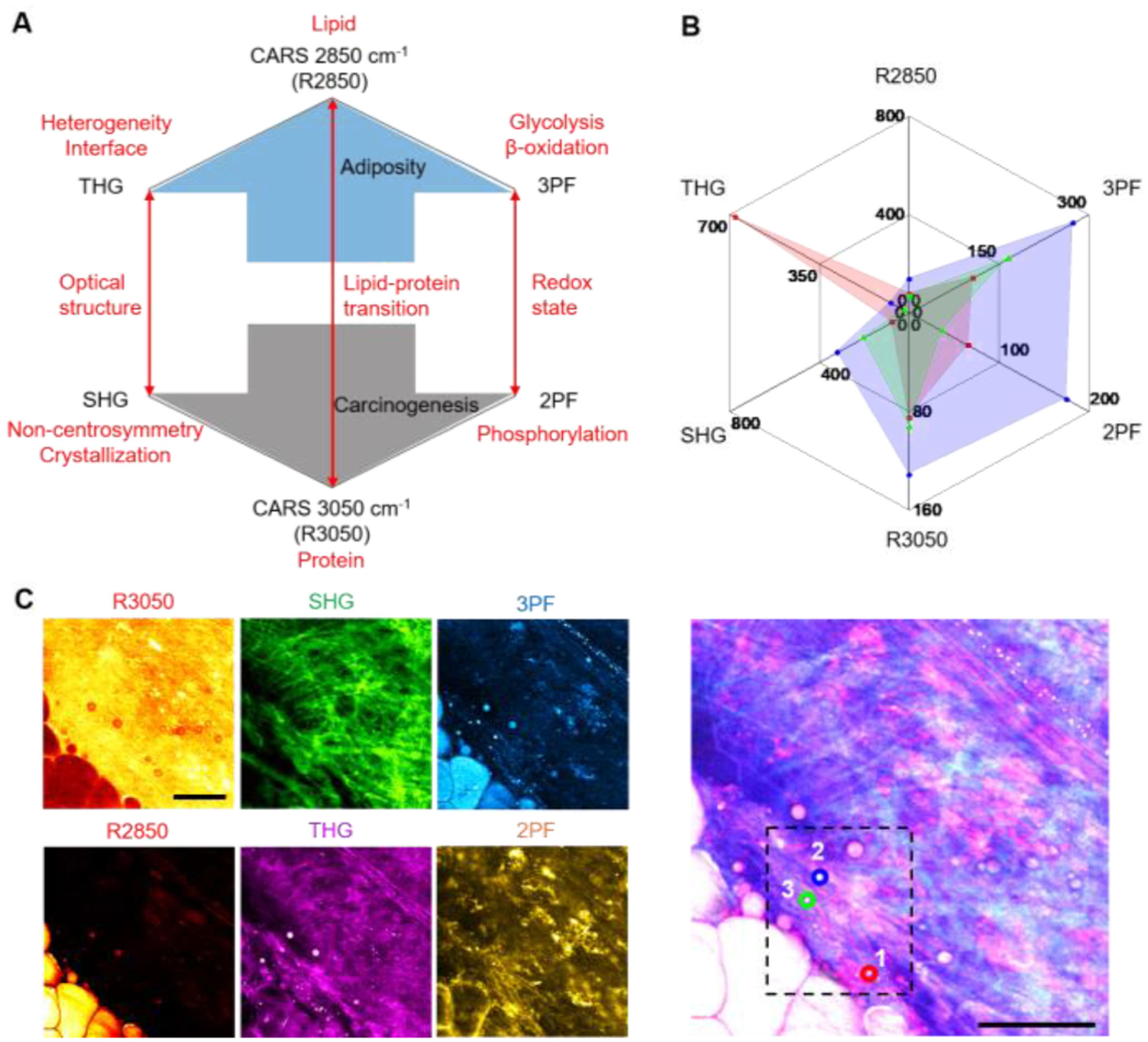
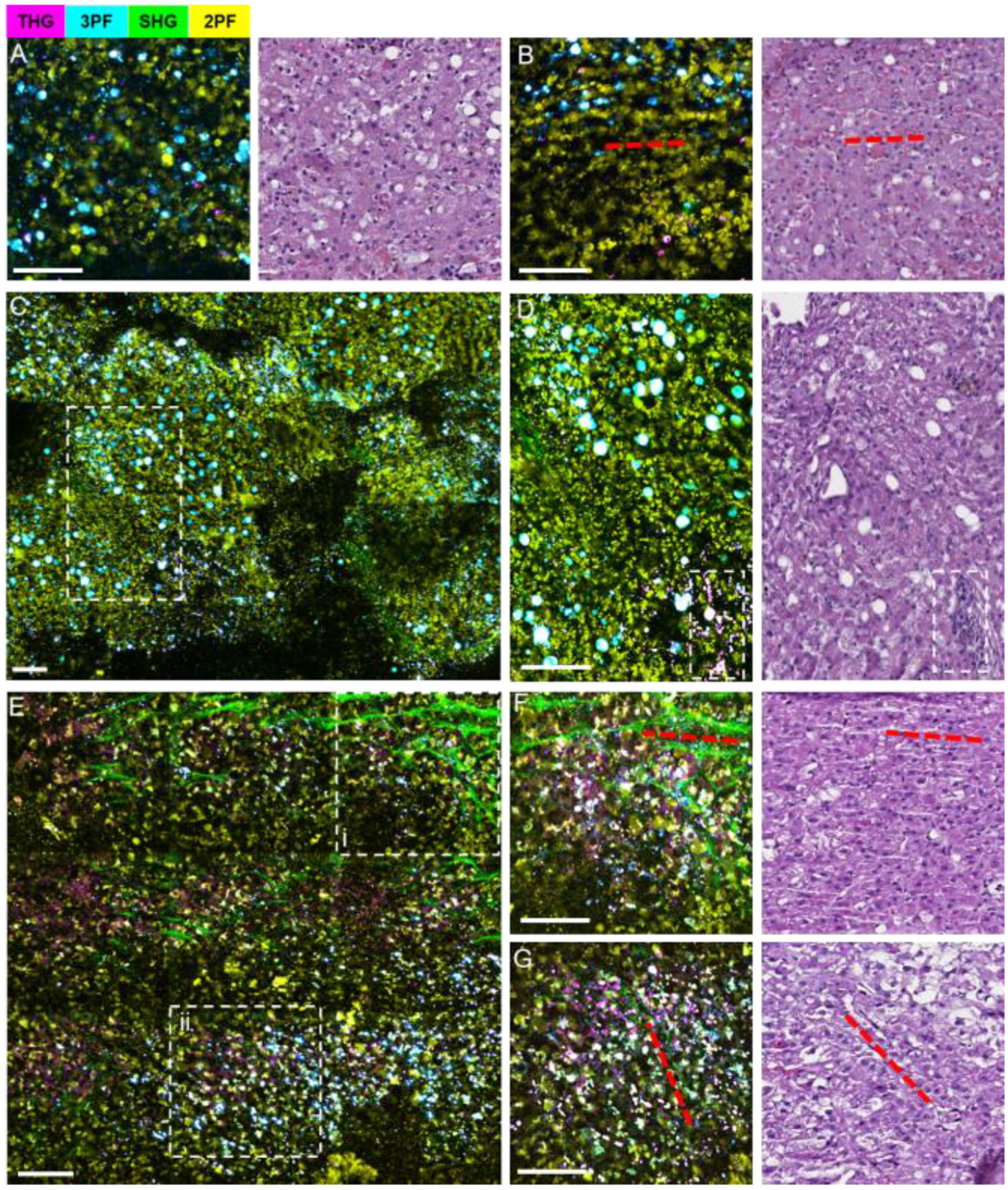
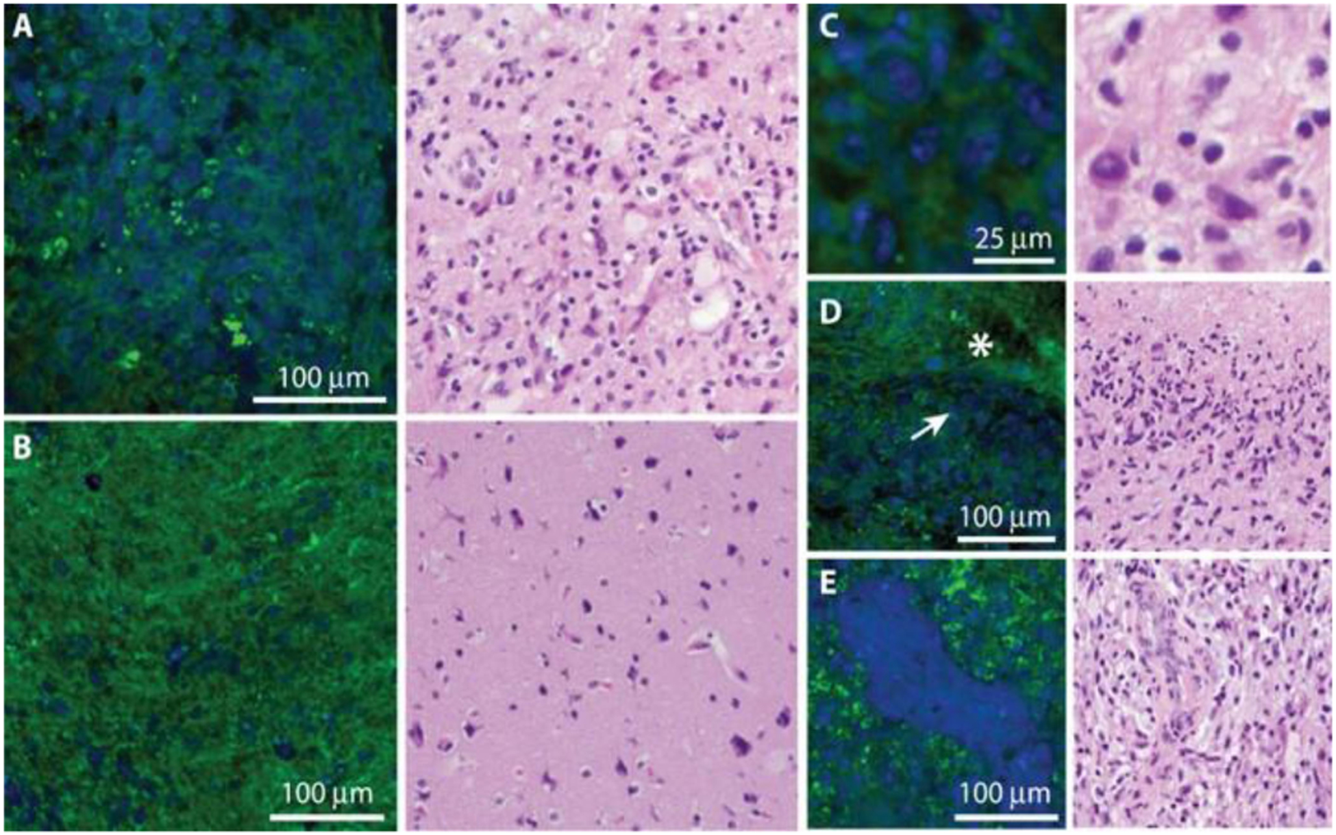
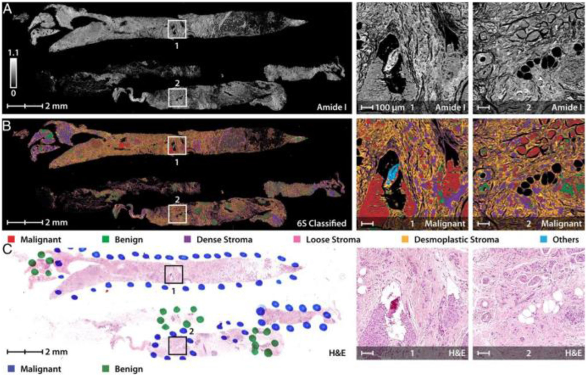
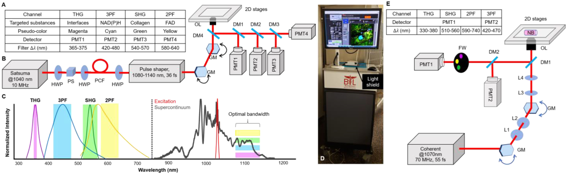
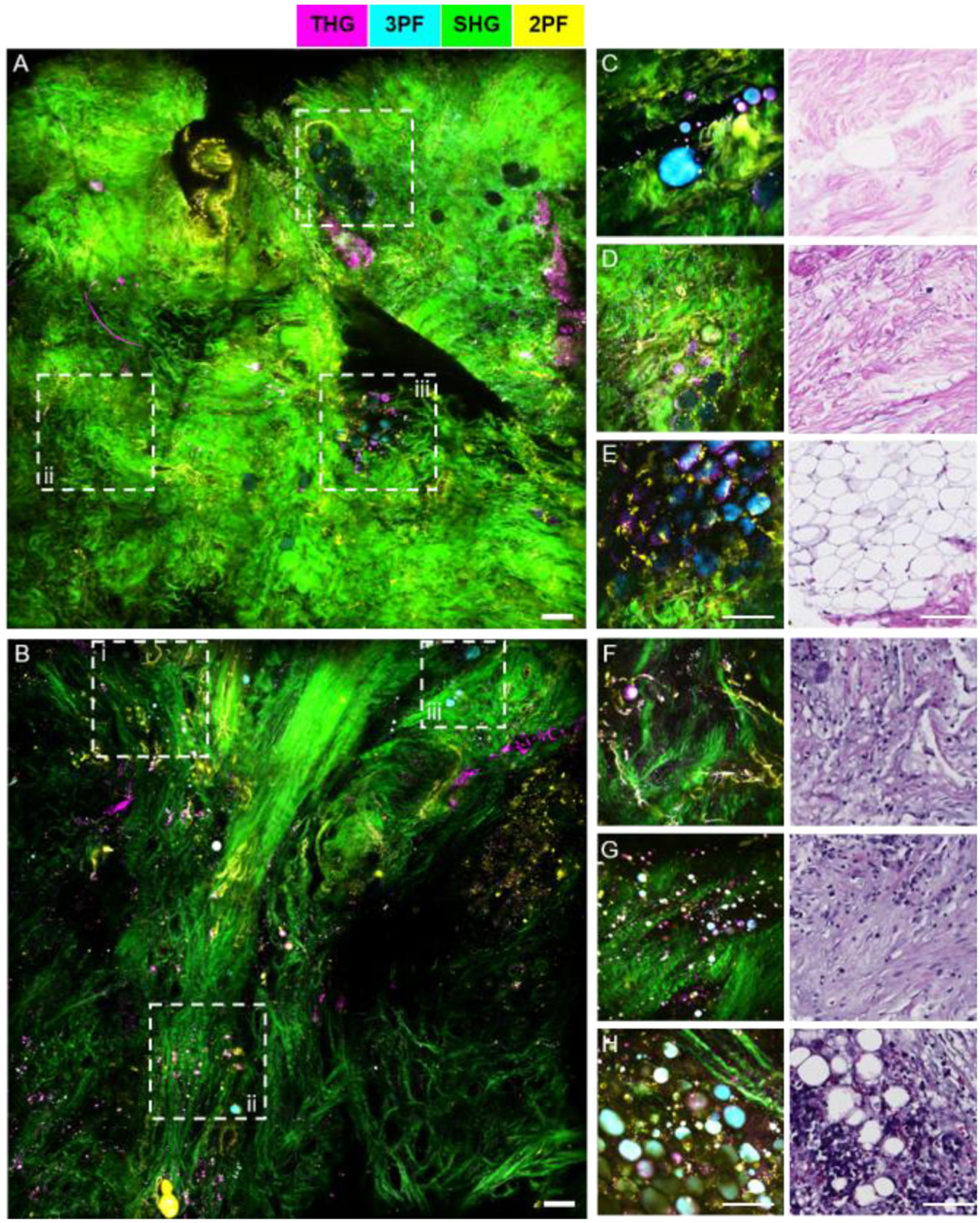
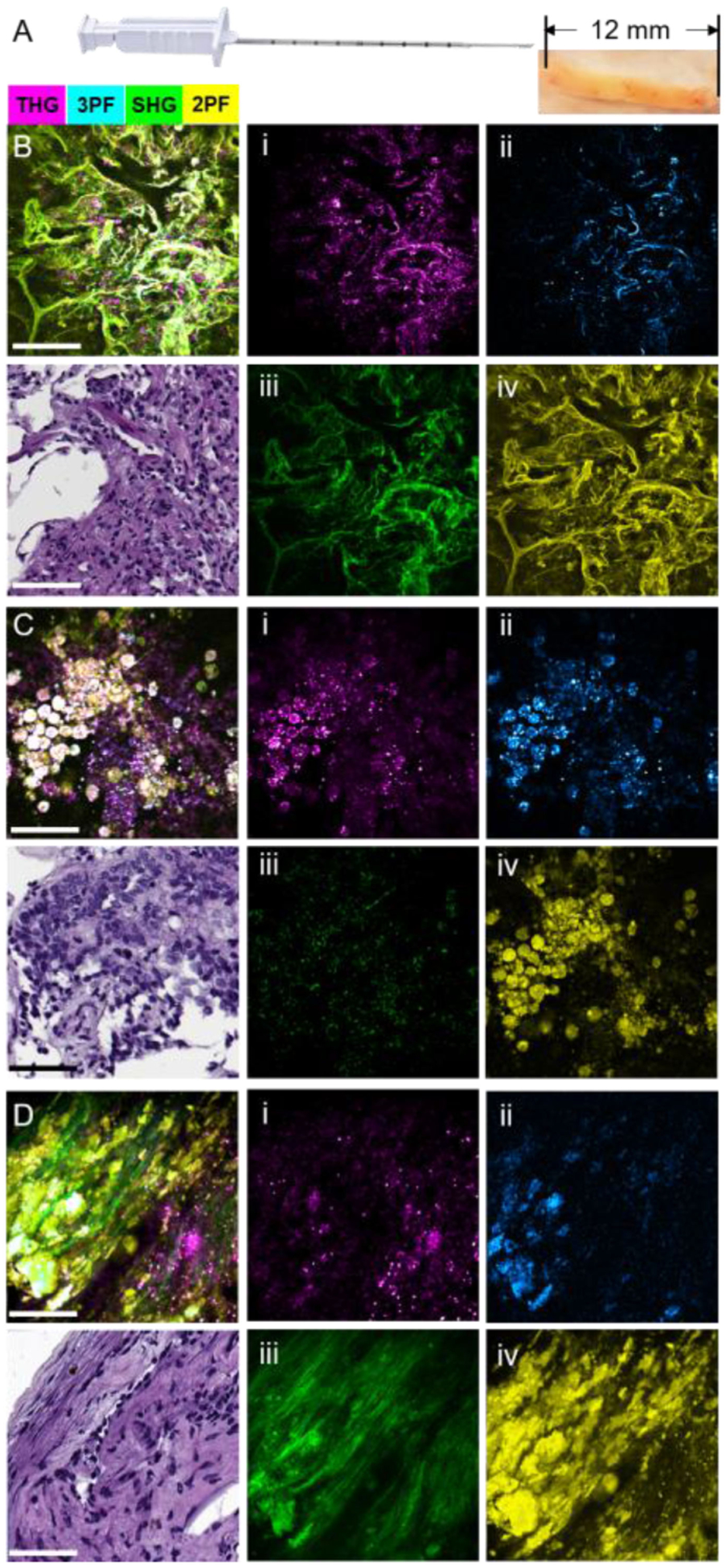
Intraoperative imaging in surgical oncology can provide information about the tumor microenvironment as well as information about the tumor margin. Visualizing microstructural features and molecular and functional dynamics may provide important diagnostic and prognostic information, especially when obtained in real-time at the point-of-procedure. A majority of current intraoperative optical techniques are based on the use of the labels, such as fluorescent dyes. However, these exogenous agents disrupt the natural microenvironment, perturb biological processes, and alter the endogenous optical signatures that cells and the microenvironment can provide. Portable nonlinear imaging systems have enabled intraoperative imaging for real-time detection and diagnosis of tissue. We review the development of a label-free multimodal nonlinear optical imaging technique that was adapted into a portable imaging system for intraoperative optical assessment of resected human breast tissue. New developments have applied this technology to assessing needle-biopsy specimens. Needle-biopsy procedures most always precede surgical resection and serve as the first sampling of suspicious masses for diagnosis. We demonstrate the diagnostic feasibility of imaging core needle-biopsy specimens during veterinary cancer surgeries. This intraoperative label-free multimodal nonlinear optical imaging technique can potentially provide a powerful tool to assist in cancer diagnosis at the point-of-procedure.
外科肿瘤学中的术中成像可以提供有关肿瘤微环境以及肿瘤边缘的信息。可视化微观结构特征以及分子和功能动态可能会提供重要的诊断和预后信息,尤其是在手术过程中实时获取这些信息时。当前大多数术中光学技术都基于使用诸如荧光染料之类的标记物。然而,这些外源性试剂会破坏自然微环境,干扰生物过程,并改变细胞和微环境所能提供的内源性光学特征。便携式非线性成像系统已实现术中成像,用于组织的实时检测和诊断。我们回顾了一种无标记多模态非线性光学成像技术的发展,该技术被应用于一种便携式成像系统,用于对切除的人类乳腺组织进行术中光学评估。新的进展已将这项技术应用于评估针吸活检标本。针吸活检程序大多在手术切除之前进行,并且作为对可疑肿块进行诊断的首次采样。我们展示了在兽医癌症手术中对粗针活检标本进行成像的诊断可行性。这种术中无标记多模态非线性光学成像技术有可能提供一个强大工具,在手术过程中协助癌症诊断。