Bai Jie, Wang Julie T-W, Rubio Noelia, Protti Andrea, Heidari Hamed, Elgogary Riham, Southern Paul, Al-Jamal Wafa' T, Sosabowski Jane, Shah Ajay M, Bals Sara, Pankhurst Quentin A, Al-Jamal Khuloud T
1. Institute of Pharmaceutical Science, Faculty of Life Sciences & Medicine, King's College London, London, SE1 9NH, UK.
2. Cardiovascular Division, James Black Centre, King's College London British Heart Foundation Centre of Excellence, London, SE5 9NU, UK.
Theranostics. 2016 Jan 1;6(3):342-56. doi: 10.7150/thno.11918. eCollection 2016.
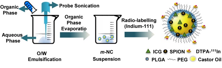
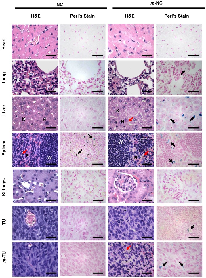
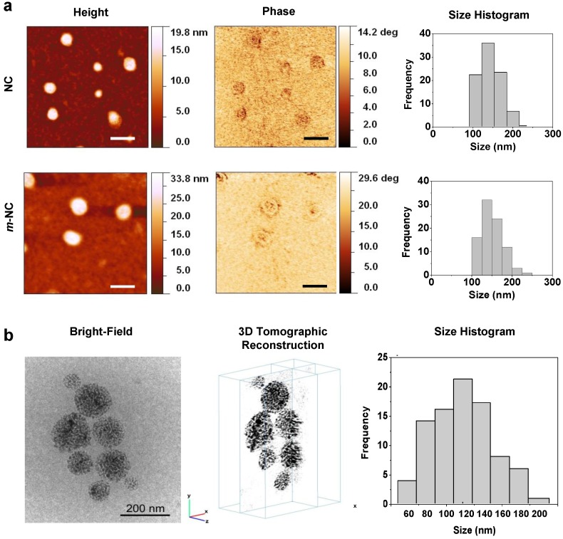
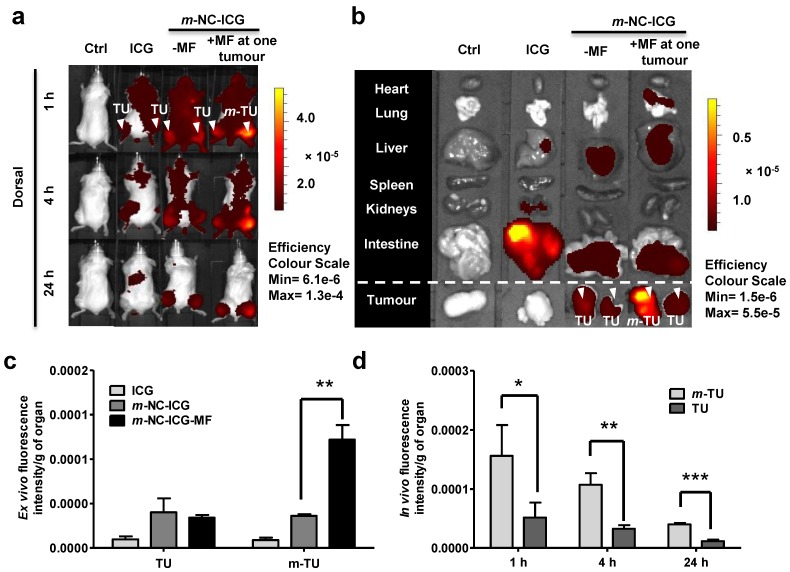
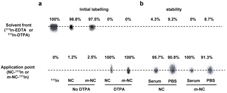
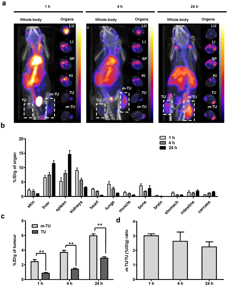
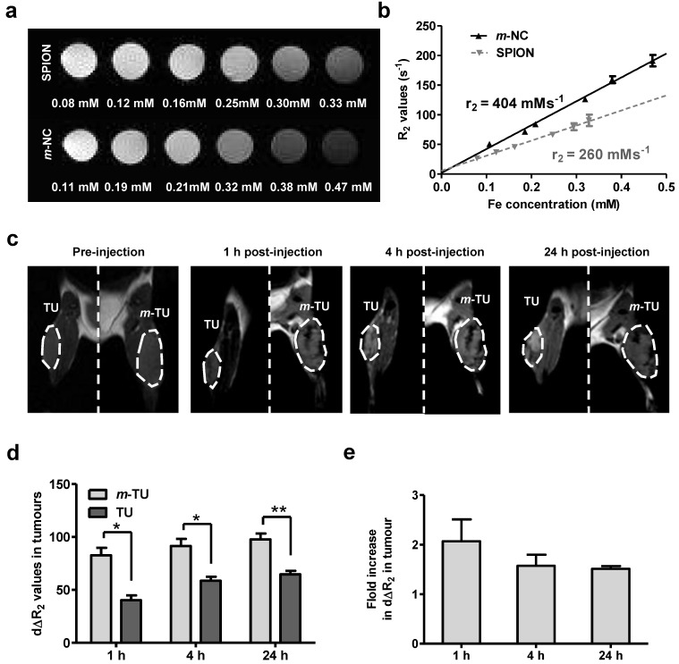
Triple-modal imaging magnetic nanocapsules, encapsulating hydrophobic superparamagnetic iron oxide nanoparticles, are formulated and used to magnetically target solid tumours after intravenous administration in tumour-bearing mice. The engineered magnetic polymeric nanocapsules m-NCs are ~200 nm in size with negative Zeta potential and shown to be spherical in shape. The loading efficiency of superparamagnetic iron oxide nanoparticles in the m-NC was ~100%. Up to ~3- and ~2.2-fold increase in tumour uptake at 1 and 24 h was achieved, when a static magnetic field was applied to the tumour for 1 hour. m-NCs, with multiple imaging probes (e.g. indocyanine green, superparamagnetic iron oxide nanoparticles and indium-111), were capable of triple-modal imaging (fluorescence/magnetic resonance/nuclear imaging) in vivo. Using triple-modal imaging is to overcome the intrinsic limitations of single modality imaging and provides complementary information on the spatial distribution of the nanocarrier within the tumour. The significant findings of this study could open up new research perspectives in using novel magnetically-responsive nanomaterials in magnetic-drug targeting combined with multi-modal imaging.
包裹疏水性超顺磁性氧化铁纳米颗粒的三模态成像磁性纳米胶囊,经制备后用于荷瘤小鼠静脉注射后对实体瘤进行磁靶向。工程化的磁性聚合物纳米胶囊m-NCs尺寸约为200 nm,具有负的zeta电位,呈球形。超顺磁性氧化铁纳米颗粒在m-NC中的负载效率约为100%。当对肿瘤施加1小时的静磁场时,在1小时和24小时时肿瘤摄取量分别增加了约3倍和2.2倍。具有多种成像探针(如吲哚菁绿、超顺磁性氧化铁纳米颗粒和铟-111)的m-NCs能够在体内进行三模态成像(荧光/磁共振/核成像)。使用三模态成像旨在克服单模态成像的固有局限性,并提供有关纳米载体在肿瘤内空间分布的补充信息。本研究的重要发现可能为在磁药物靶向结合多模态成像中使用新型磁响应纳米材料开辟新的研究前景。