Perrod Guillaume, Rahmi Gabriel, Pidial Laetitia, Camilleri Sophie, Bellucci Alexandre, Casanova Amaury, Viel Thomas, Tavitian Bertrand, Cellier Christophe, Clement Olivier
Université Paris Descartes Sorbonne Paris cité, Assistance Publique-Hôpitaux de Paris, Department of Gastroenterology, Hôpital Européen Georges Pompidou, 20 rue Leblanc, 75015 Paris, France.
Université Paris Descartes Sorbonne Paris cité, Laboratoire imagerie de l'angiogenèse et plateforme d'imagerie du petit animal, UMR-S970, 56 rue Leblanc, 75015 Paris, France.
PLoS One. 2016 Mar 1;11(3):e0148249. doi: 10.1371/journal.pone.0148249. eCollection 2016.
BACKGROUND & AIMS: Extended esophageal endoscopic submucosal dissection (ESD) is highly responsible for esophageal stricture. We conducted a comparative study in a porcine model to evaluate the effectiveness of adipose tissue-derived stromal cell (ADSC) double cell sheet transplantation.
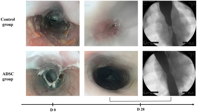
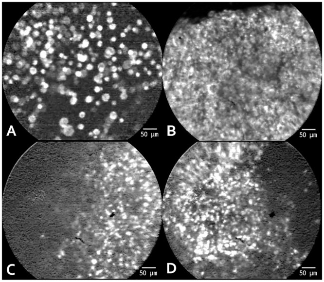
Twelve female pigs were treated with 5 cm long hemi-circumferential ESD and randomized in two groups. ADSC group (n = 6) received 4 double cell sheets of allogenic ADSC on a paper support membrane and control group (n = 6) received 4 paper support membranes. ADSC were labelled with PKH-67 fluorophore to allow probe-based confocal laser endomicroscopie (pCLE) monitoring. After 28 days follow-up, animals were sacrificed. At days 3, 14 and 28, endoscopic evaluation with pCLE and esophagography were performed.
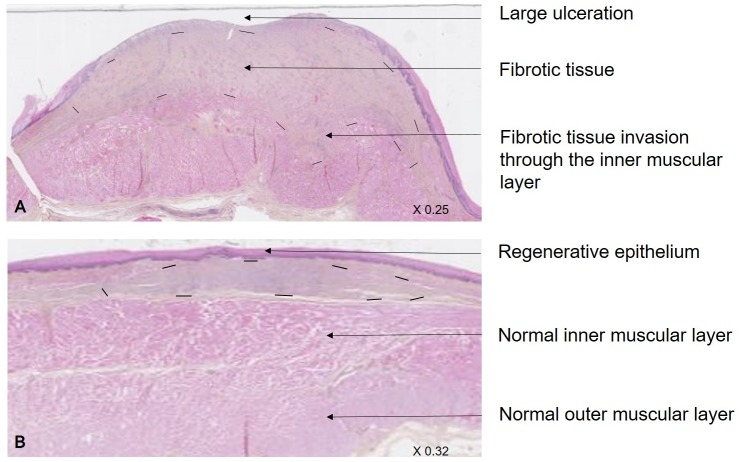
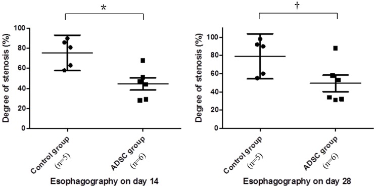
One animal from the control group was excluded (anesthetic complication). Animals from ADSC group showed less frequent alimentary trouble (17% vs 80%; P = 0.08) and higher gain weight on day 28. pCLE demonstrated a compatible cell signal in 4 animals of the ADSC group at day 3. In ADSC group, endoscopy showed that 1 out of 6 (17%) animals developed a severe esophageal stricture comparatively to 100% (5/5) in the control group; P = 0.015. Esophagography demonstrated a decreased degree of stricture in the ADSC group on day 14 (44% vs 81%; P = 0.017) and day 28 (46% vs 90%; P = 0.035). Histological analysis showed a decreased fibrosis development in the ADSC group, in terms of surface (9.7 vs 26.1 mm²; P = 0.017) and maximal depth (1.6 vs 3.2 mm; P = 0.052).
In this model, transplantation of allogenic ADSC organized in double cell sheets after extended esophegeal ESD is strongly associated with a lower esophageal stricture's rate.
扩大食管内镜黏膜下剥离术(ESD)导致食管狭窄的风险很高。我们在猪模型中进行了一项比较研究,以评估脂肪组织来源的基质细胞(ADSC)双层细胞片移植的有效性。
12只雌性猪接受5厘米长的半周向ESD治疗,并随机分为两组。ADSC组(n = 6)在纸质支撑膜上接受4片同种异体ADSC双层细胞片,对照组(n = 6)接受4片纸质支撑膜。用PKH-67荧光团标记ADSC,以便进行基于探针的共聚焦激光内镜显微镜检查(pCLE)监测。随访28天后,处死动物。在第3、14和28天,进行pCLE内镜评估和食管造影。
对照组有1只动物被排除(麻醉并发症)。ADSC组动物出现消化问题的频率较低(17%对80%;P = 0.08),且在第28天体重增加更多。pCLE显示,ADSC组4只动物在第3天有相容的细胞信号。在ADSC组,内镜检查显示,6只动物中有1只(17%)出现严重食管狭窄,而对照组为100%(5/5);P = 0.015。食管造影显示,ADSC组在第14天(44%对81%;P = 0.017)和第28天(46%对90%;P = 0.035)狭窄程度降低。组织学分析显示,ADSC组在表面(9.7对26.1平方毫米;P = 0.017)和最大深度(1.6对3.2毫米;P = 0.052)方面纤维化发展减少。
在该模型中,扩大食管ESD后移植组织成双层细胞片的同种异体ADSC与较低的食管狭窄率密切相关。