• 文献检索
  • 文档翻译
  • 深度研究
  • 学术资讯
  • Suppr Zotero 插件Zotero 插件
  • 邀请有礼
  • 套餐&价格
  • 历史记录
应用&插件
Suppr Zotero 插件Zotero 插件浏览器插件Mac 客户端Windows 客户端微信小程序
定价
高级版会员购买积分包购买API积分包
服务
文献检索文档翻译深度研究API 文档MCP 服务
关于我们
关于 Suppr公司介绍联系我们用户协议隐私条款
关注我们

Suppr 超能文献

核心技术专利:CN118964589B侵权必究
粤ICP备2023148730 号-1Suppr @ 2026

文献检索

告别复杂PubMed语法,用中文像聊天一样搜索,搜遍4000万医学文献。AI智能推荐,让科研检索更轻松。

立即免费搜索

文件翻译

保留排版,准确专业,支持PDF/Word/PPT等文件格式,支持 12+语言互译。

免费翻译文档

深度研究

AI帮你快速写综述,25分钟生成高质量综述,智能提取关键信息,辅助科研写作。

立即免费体验

使用特温特光声乳腺镜进行乳腺成像的最新技术:对恶性肿瘤进行31次测量的结果

The state of the art in breast imaging using the Twente Photoacoustic Mammoscope: results from 31 measurements on malignancies.

作者信息

Heijblom Michelle, Piras Daniele, van den Engh Frank M, van der Schaaf Margreet, Klaase Joost M, Steenbergen Wiendelt, Manohar Srirang

机构信息

Biomedical Photonic Imaging Group, MIRA Institute for Biomedical Technology and Technical Medicine, University of Twente, P.O. Box 217, 7500 AE, Enschede, The Netherlands.

Center for Breast Care, Medisch Spectrum Twente, P.O. Box 50.000, 7500 KA, Enschede, The Netherlands.

出版信息

Eur Radiol. 2016 Nov;26(11):3874-3887. doi: 10.1007/s00330-016-4240-7. Epub 2016 Mar 5.

DOI:10.1007/s00330-016-4240-7
PMID:26945762
原文链接:https://pmc.ncbi.nlm.nih.gov/articles/PMC5052314/
Abstract

OBJECTIVES

Photoacoustic mammography is potentially an ideal technique, however, the amount of patient data is limited. To further our understanding of the in vivo performance of the method and to guide further research and development, we imaged 33 breast malignancies using the research system - the Twente Photoacoustic Mammoscope (PAM).

METHODS

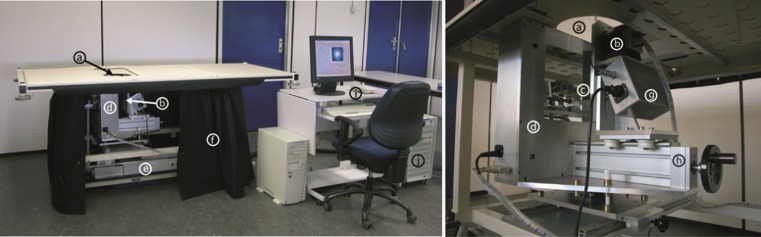
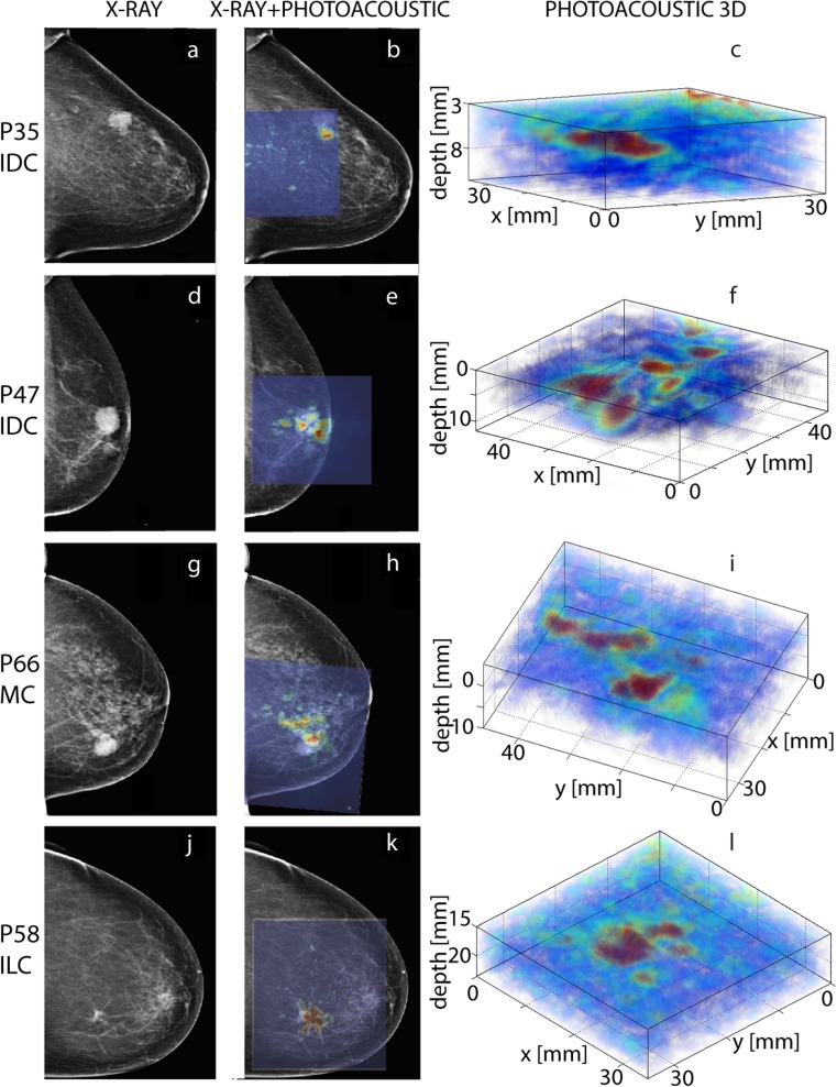
Thirty-one patients participated in this retrospective, observational study. The study and informed consent procedure were approved by the local ethics committee. PAM uses 1,064 nm light for excitation with a planar, 588-element, 1-MHz ultrasound array for detection. Photoacoustic lesion visibility and appearance were compared with conventional imaging (x-ray mammography and ultrasonography) findings, histopathology and patient demographics.

RESULTS

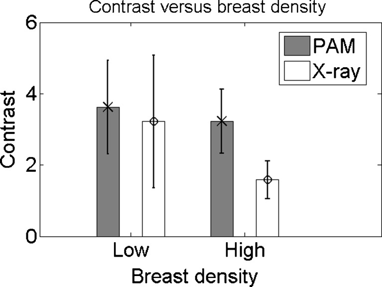
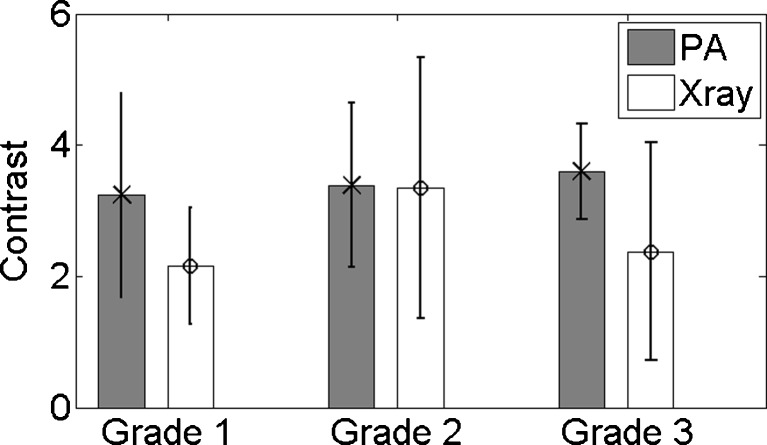
Of 33 malignancies 32 were visualized with high contrast and good co-localization with conventional imaging. The contrast of the detected malignancies was independent of radiographic breast density, and size estimation was reasonably good with an average 28 % deviation from histology. However, the presence of contrast areas outside the malignant region is suggestive for low specificity of the current system. Statistical analyses did not reveal any further relationship between PAM results and patient demographics nor lesion characteristics.

CONCLUSIONS

The results confirm the high potential of photoacoustic mammography in future breast care.

KEY POINTS

• Photoacoustic breast imaging visualizes malignancies with high imaging contrast. • Photoacoustic lesion contrast is independent of the mammographically estimated breast density. • No clear relationship exists between photoacoustic characteristics and lesion type, grade, etc. • Photoacoustic specificity to breast cancer from some cases is not yet optimal.

摘要

目的

光声乳腺成像可能是一种理想的技术,然而,患者数据量有限。为了进一步了解该方法的体内性能并指导进一步的研究与开发,我们使用研究系统——特温特光声乳腺镜(PAM)对33例乳腺恶性肿瘤进行了成像。

方法

31名患者参与了这项回顾性观察研究。该研究及知情同意程序获得了当地伦理委员会的批准。PAM使用1064纳米的光进行激发,采用平面588阵元、1兆赫的超声阵列进行检测。将光声病变的可见性和表现与传统成像(乳腺X线摄影和超声检查)结果、组织病理学及患者人口统计学数据进行比较。

结果

33例恶性肿瘤中有32例在光声成像中显示出高对比度,且与传统成像具有良好的共定位。检测到的恶性肿瘤的对比度与乳腺X线摄影密度无关,大小估计相当准确,与组织学结果的平均偏差为28%。然而,恶性区域外存在对比区域提示当前系统的特异性较低。统计分析未发现PAM结果与患者人口统计学数据或病变特征之间存在任何进一步的关系。

结论

结果证实了光声乳腺成像在未来乳腺护理中的巨大潜力。

关键点

• 光声乳腺成像能够以高成像对比度显示恶性肿瘤。• 光声病变对比度与乳腺X线摄影估计的乳腺密度无关。• 光声特征与病变类型、分级等之间不存在明确关系。• 某些病例中光声对乳腺癌的特异性尚未达到最佳。

https://cdn.ncbi.nlm.nih.gov/pmc/blobs/0676/5052314/9362eaa59a07/330_2016_4240_Fig4_HTML.jpg
https://cdn.ncbi.nlm.nih.gov/pmc/blobs/0676/5052314/4610bc17e58d/330_2016_4240_Fig1_HTML.jpg
https://cdn.ncbi.nlm.nih.gov/pmc/blobs/0676/5052314/214a56504c1f/330_2016_4240_Fig2_HTML.jpg
https://cdn.ncbi.nlm.nih.gov/pmc/blobs/0676/5052314/298e255ae7fb/330_2016_4240_Fig3_HTML.jpg
https://cdn.ncbi.nlm.nih.gov/pmc/blobs/0676/5052314/9362eaa59a07/330_2016_4240_Fig4_HTML.jpg

相似文献

1
The state of the art in breast imaging using the Twente Photoacoustic Mammoscope: results from 31 measurements on malignancies.使用特温特光声乳腺镜进行乳腺成像的最新技术:对恶性肿瘤进行31次测量的结果
Eur Radiol. 2016 Nov;26(11):3874-3887. doi: 10.1007/s00330-016-4240-7. Epub 2016 Mar 5.
2
Visualizing breast cancer using the Twente photoacoustic mammoscope: what do we learn from twelve new patient measurements?使用特温特光声乳腺镜可视化乳腺癌:我们从十二例新患者测量中了解到了什么?
Opt Express. 2012 May 21;20(11):11582-97. doi: 10.1364/OE.20.011582.
3
Appearance of breast cysts in planar geometry photoacoustic mammography using 1064-nm excitation.在使用 1064nm 激发光的平面几何光声乳腺成像中出现的乳腺囊肿。
J Biomed Opt. 2013 Dec;18(12):126009. doi: 10.1117/1.JBO.18.12.126009.
4
Twente Photoacoustic Mammoscope 2: system overview and three-dimensional vascular network images in healthy breasts.特文特光声乳腺成像仪 2 型:系统概述及健康乳房的三维血管网络图像。
J Biomed Opt. 2019 Oct;24(12):1-12. doi: 10.1117/1.JBO.24.12.121909.
5
Photoacoustic mammography: initial clinical results.光声乳腺成像:初步临床结果。
Breast Cancer. 2014 Mar;21(2):146-53. doi: 10.1007/s12282-012-0363-0. Epub 2012 Apr 7.
6
Photoacoustic image patterns of breast carcinoma and comparisons with Magnetic Resonance Imaging and vascular stained histopathology.乳腺癌的光声图像模式及其与磁共振成像和血管染色组织病理学的比较。
Sci Rep. 2015 Jul 10;5:11778. doi: 10.1038/srep11778.
7
Imaging breast malignancies with the Twente Photoacoustic Mammoscope 2.用 Twente 光声乳腺影像仪 2 号对乳腺恶性肿瘤进行成像。
PLoS One. 2023 Mar 2;18(3):e0281434. doi: 10.1371/journal.pone.0281434. eCollection 2023.
8
Photoacoustic breast tomography prototypes with reported human applications.已报道有人类应用的光声乳腺断层成像原型。
Eur Radiol. 2015 Aug;25(8):2205-13. doi: 10.1007/s00330-015-3647-x. Epub 2015 Feb 27.
9
Combined photoacoustic and acoustic imaging of human breast specimens in the mammographic geometry.在乳腺摄影几何形状下对人体乳腺标本进行的光声和声学联合成像。
Ultrasound Med Biol. 2013 Nov;39(11):2176-84. doi: 10.1016/j.ultrasmedbio.2013.05.018. Epub 2013 Aug 22.
10
Clinical photoacoustic breast imaging: the Twente experience.临床光声乳腺成像:特温特大学的经验。
IEEE Pulse. 2015 May-Jun;6(3):42-6. doi: 10.1109/MPUL.2015.2409102.

引用本文的文献

1
Resolution Enhancement Strategies in Photoacoustic Microscopy: A Comprehensive Review.光声显微镜中的分辨率增强策略:全面综述
Micromachines (Basel). 2024 Nov 30;15(12):1463. doi: 10.3390/mi15121463.
2
Optical Breast Imaging: A Review of Physical Principles, Technologies, and Clinical Applications.光学乳腺成像:物理原理、技术及临床应用综述
J Breast Imaging. 2023 Sep-Oct;5(5):520-537. doi: 10.1093/jbi/wbad057. Epub 2023 Sep 22.
3
Breast Cancer Diagnosis Using Extended-Wavelength-Diffuse Reflectance Spectroscopy (EW-DRS)-Proof of Concept in Ex Vivo Breast Specimens Using Machine Learning.

本文引用的文献

1
High resolution functional photoacoustic tomography of breast cancer.乳腺癌的高分辨率功能光声断层成像
Med Phys. 2015 Sep;42(9):5321-8. doi: 10.1118/1.4928598.
2
Photoacoustic image patterns of breast carcinoma and comparisons with Magnetic Resonance Imaging and vascular stained histopathology.乳腺癌的光声图像模式及其与磁共振成像和血管染色组织病理学的比较。
Sci Rep. 2015 Jul 10;5:11778. doi: 10.1038/srep11778.
3
Clinical photoacoustic breast imaging: the Twente experience.临床光声乳腺成像:特温特大学的经验。
使用扩展波长漫反射光谱法(EW-DRS)诊断乳腺癌——基于机器学习的离体乳腺标本概念验证
Diagnostics (Basel). 2023 Sep 28;13(19):3076. doi: 10.3390/diagnostics13193076.
4
Biomarkers of aging.衰老的生物标志物。
Sci China Life Sci. 2023 May;66(5):893-1066. doi: 10.1007/s11427-023-2305-0. Epub 2023 Apr 11.
5
The application of optical technology in the diagnosis and therapy of oxidative stress-mediated hepatic ischemia-reperfusion injury.光学技术在氧化应激介导的肝脏缺血再灌注损伤诊断与治疗中的应用。
Front Bioeng Biotechnol. 2023 Feb 20;11:1133039. doi: 10.3389/fbioe.2023.1133039. eCollection 2023.
6
Imaging breast malignancies with the Twente Photoacoustic Mammoscope 2.用 Twente 光声乳腺影像仪 2 号对乳腺恶性肿瘤进行成像。
PLoS One. 2023 Mar 2;18(3):e0281434. doi: 10.1371/journal.pone.0281434. eCollection 2023.
7
A Comprehensive Review on Photoacoustic-Based Devices for Biomedical Applications.基于光声的生物医学应用设备的综合评述
Sensors (Basel). 2022 Dec 6;22(23):9541. doi: 10.3390/s22239541.
8
An optoacoustic imaging feature set to characterise blood vessels surrounding benign and malignant breast lesions.一种用于表征良性和恶性乳腺病变周围血管的光声成像特征集。
Photoacoustics. 2022 Jul 4;27:100383. doi: 10.1016/j.pacs.2022.100383. eCollection 2022 Sep.
9
The emerging role of photoacoustic imaging in clinical oncology.光声成像在临床肿瘤学中的新兴作用。
Nat Rev Clin Oncol. 2022 Jun;19(6):365-384. doi: 10.1038/s41571-022-00615-3. Epub 2022 Mar 23.
10
The Potential of Photoacoustic Imaging in Radiation Oncology.光声成像在放射肿瘤学中的潜力
Front Oncol. 2022 Mar 3;12:803777. doi: 10.3389/fonc.2022.803777. eCollection 2022.
IEEE Pulse. 2015 May-Jun;6(3):42-6. doi: 10.1109/MPUL.2015.2409102.
4
Mesoscopic and macroscopic optoacoustic imaging of cancer.癌症的介观和宏观光声成像。
Cancer Res. 2015 Apr 15;75(8):1548-59. doi: 10.1158/0008-5472.CAN-14-2522. Epub 2015 Apr 2.
5
Hemodynamic signature of breast cancer under fractional mammographic compression using a dynamic diffuse optical tomography system.使用动态漫射光学层析成像系统在乳腺X线摄影部分压迫下乳腺癌的血流动力学特征。
Biomed Opt Express. 2013 Nov 22;4(12):2911-24. doi: 10.1364/BOE.4.002911. eCollection 2013.
6
Appearance of breast cysts in planar geometry photoacoustic mammography using 1064-nm excitation.在使用 1064nm 激发光的平面几何光声乳腺成像中出现的乳腺囊肿。
J Biomed Opt. 2013 Dec;18(12):126009. doi: 10.1117/1.JBO.18.12.126009.
7
Dedicated 3D photoacoustic breast imaging.专用 3D 光声乳腺成像。
Med Phys. 2013 Nov;40(11):113301. doi: 10.1118/1.4824317.
8
Optoacoustic imaging and tomography: reconstruction approaches and outstanding challenges in image performance and quantification.光声成像和层析成像:在图像性能和定量方面的重建方法及突出挑战。
Sensors (Basel). 2013 Jun 4;13(6):7345-84. doi: 10.3390/s130607345.
9
Novel methods and tracers for breast cancer imaging.用于乳腺癌成像的新方法和示踪剂。
Semin Nucl Med. 2013 Jul;43(4):324-9. doi: 10.1053/j.semnuclmed.2013.02.003.
10
Beyond mammography: new frontiers in breast cancer screening.超越乳腺 X 光检查:乳腺癌筛查的新领域。
Am J Med. 2013 Jun;126(6):472-9. doi: 10.1016/j.amjmed.2012.11.025. Epub 2013 Apr 3.