den Haan Jurre, Janssen Sarah F, van de Kreeke Jacoba A, Scheltens Philip, Verbraak Frank D, Bouwman Femke H
Department of Neurology, Alzheimer Center, VU University Medical Center, Amsterdam, The Netherlands.
Ophthalmology Department, VU University Medical Center, Amsterdam, The Netherlands.
Alzheimers Dement (Amst). 2017 Nov 6;10:49-55. doi: 10.1016/j.dadm.2017.10.005. eCollection 2018.
The retina may reflect Alzheimer's disease (AD) neuropathological changes and is easily visualized with optical coherence tomography (OCT). Retinal thickness decrease has been correlated to AD, however, without information on amyloid status. We correlated retinal (layer) thickness to AD biomarkers in amyloid-positive early-onset AD (EOAD) patients and amyloid-negative controls.
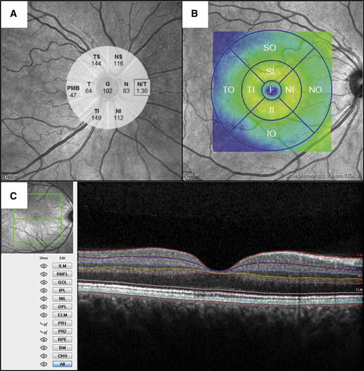
We measured macular thickness and peripapillary retinal nerve fiber layer thickness with OCT in 15 EOAD patients and 15 controls and correlated retinal thickness to visual rating scores for atrophy on magnetic resonance imaging.
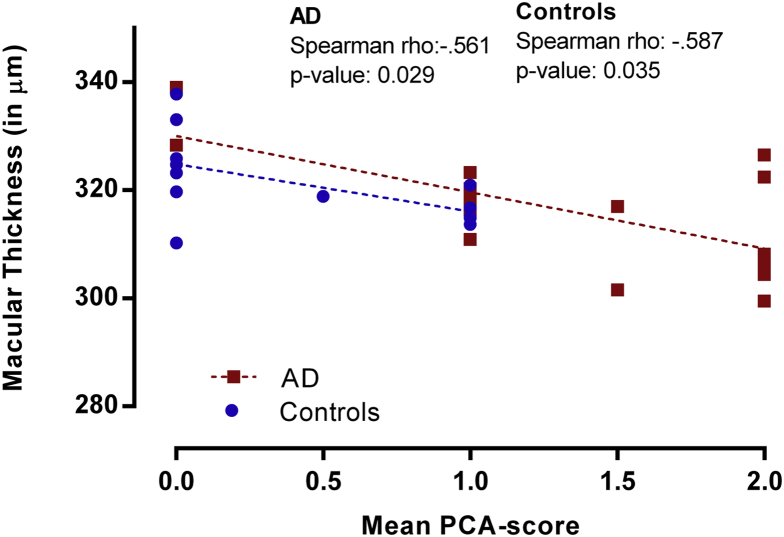
Total macular thickness correlated to parietal cortical atrophy in both groups (Spearman ρ -0.603, = .001). Macular and peripapillary retinal nerve fiber layer thicknesses were not significantly decreased in EOAD compared to controls.
Retinal thickness does not discriminate EOAD from controls but is correlated to parietal cortical atrophy in both groups. These findings may suggest reflection of cerebral cortical changes in the retina, independent of amyloid.
视网膜可能反映阿尔茨海默病(AD)的神经病理变化,并且通过光学相干断层扫描(OCT)很容易观察到。视网膜厚度降低与AD相关,但未涉及淀粉样蛋白状态的信息。我们将视网膜(各层)厚度与淀粉样蛋白阳性早发型AD(EOAD)患者及淀粉样蛋白阴性对照者的AD生物标志物进行了关联分析。
我们用OCT测量了15例EOAD患者和15例对照者的黄斑厚度及视乳头周围视网膜神经纤维层厚度,并将视网膜厚度与磁共振成像上萎缩的视觉评分进行关联分析。
两组中,黄斑总厚度均与顶叶皮质萎缩相关(斯皮尔曼ρ=-0.603,P=0.001)。与对照者相比,EOAD患者的黄斑和视乳头周围视网膜神经纤维层厚度未显著降低。
视网膜厚度不能区分EOAD患者与对照者,但两组中视网膜厚度均与顶叶皮质萎缩相关。这些发现可能提示视网膜中脑皮质变化的反映,与淀粉样蛋白无关。