Vasquez Erick S, Feugang Jean M, Willard Scott T, Ryan Peter L, Walters Keisha B
Department of Chemical and Materials Engineering, University of Dayton, Dayton, OH, 45469, USA.
Facility for Cellular and Organismal Imaging, Mississippi State University, Mississippi State, MS, 39762, USA.
J Nanobiotechnology. 2016 Mar 17;14:20. doi: 10.1186/s12951-016-0168-y.
Nanoparticles have emerged as key materials for developing applications in nanomedicine, nanobiotechnology, bioimaging and theranostics. Existing bioimaging technologies include bioluminescent resonance energy transfer-conjugated quantum dots (BRET-QDs). Despite the current use of BRET-QDs for bioimaging, there are strong concerns about QD nanocomposites containing cadmium which exhibits potential cellular toxicity.
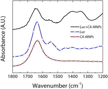
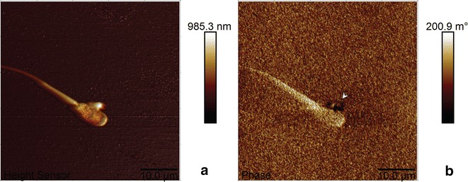
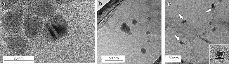
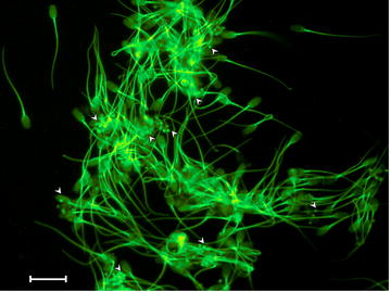
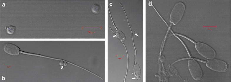
In this study, bioluminescent composites comprised of magnetic nanoparticles and firefly luciferase (Photinus pyralis) are examined as potential light-emitting agents for imaging, detection, and tracking mammalian spermatozoa. Characterization was carried out using infrared spectroscopy, TEM and cryo-TEM imaging, and ζ-potential measurements to demonstrate the successful preparation of these nanocomposites. Binding interactions between the synthesized nanoparticles and spermatozoon were characterized using confocal and atomic/magnetic force microscopy. Bioluminescence imaging and UV-visible-NIR microscopy results showed light emission from sperm samples incubated with the firefly luciferase-modified nanoparticles. Therefore, these newly synthesized luciferase-modified magnetic nanoparticles show promise as substitutes for QD labeling, and can potentially also be used for in vivo manipulation and tracking, as well as MRI techniques.
These preliminary data indicate that luciferase-magnetic nanoparticle composites can potentially be used for spermatozoa detection and imaging. Their magnetic properties add additional functionality to allow for manipulation, sorting, or tracking of cells using magnetic techniques.
纳米颗粒已成为纳米医学、纳米生物技术、生物成像和治疗诊断学等领域开发应用的关键材料。现有的生物成像技术包括生物发光共振能量转移共轭量子点(BRET-QDs)。尽管目前BRET-QDs用于生物成像,但人们对含镉的量子点纳米复合材料深感担忧,因为镉具有潜在的细胞毒性。
在本研究中,由磁性纳米颗粒和萤火虫荧光素酶(Photinus pyralis)组成的生物发光复合材料被作为成像、检测和追踪哺乳动物精子的潜在发光剂进行了研究。使用红外光谱、透射电子显微镜(TEM)和低温TEM成像以及ζ电位测量进行表征,以证明这些纳米复合材料的成功制备。使用共聚焦显微镜以及原子力显微镜/磁力显微镜对合成的纳米颗粒与精子之间的结合相互作用进行了表征。生物发光成像和紫外-可见-近红外显微镜结果显示,与萤火虫荧光素酶修饰的纳米颗粒孵育的精子样本发出了光。因此,这些新合成的荧光素酶修饰的磁性纳米颗粒有望替代量子点标记,并有可能用于体内操作和追踪以及磁共振成像(MRI)技术。
这些初步数据表明,荧光素酶-磁性纳米颗粒复合材料有潜力用于精子检测和成像。它们的磁性增加了额外的功能,能够使用磁性技术对细胞进行操作、分选或追踪。