• 文献检索
  • 文档翻译
  • 深度研究
  • 学术资讯
  • Suppr Zotero 插件Zotero 插件
  • 邀请有礼
  • 套餐&价格
  • 历史记录
应用&插件
Suppr Zotero 插件Zotero 插件浏览器插件Mac 客户端Windows 客户端微信小程序
定价
高级版会员购买积分包购买API积分包
服务
文献检索文档翻译深度研究API 文档MCP 服务
关于我们
关于 Suppr公司介绍联系我们用户协议隐私条款
关注我们

Suppr 超能文献

核心技术专利:CN118964589B侵权必究
粤ICP备2023148730 号-1Suppr @ 2026

文献检索

告别复杂PubMed语法,用中文像聊天一样搜索,搜遍4000万医学文献。AI智能推荐,让科研检索更轻松。

立即免费搜索

文件翻译

保留排版,准确专业,支持PDF/Word/PPT等文件格式,支持 12+语言互译。

免费翻译文档

深度研究

AI帮你快速写综述,25分钟生成高质量综述,智能提取关键信息,辅助科研写作。

立即免费体验

通过X射线相衬断层扫描对脂质、蛋白质和水含量进行定量三维成像

Quantitative Three-Dimensional Imaging of Lipid, Protein, and Water Contents via X-Ray Phase-Contrast Tomography.

作者信息

Willner Marian, Viermetz Manuel, Marschner Mathias, Scherer Kai, Braun Christian, Fingerle Alexander, Noël Peter, Rummeny Ernst, Pfeiffer Franz, Herzen Julia

机构信息

Department of Physics and Institute of Medical Engineering, Technische Universität München, Garching, Germany.

Department of Diagnostic and Interventional Radiology, Technische Universität München, Munich, Germany.

出版信息

PLoS One. 2016 Mar 22;11(3):e0151889. doi: 10.1371/journal.pone.0151889. eCollection 2016.

DOI:10.1371/journal.pone.0151889
PMID:27003308
原文链接:https://pmc.ncbi.nlm.nih.gov/articles/PMC4803315/
Abstract

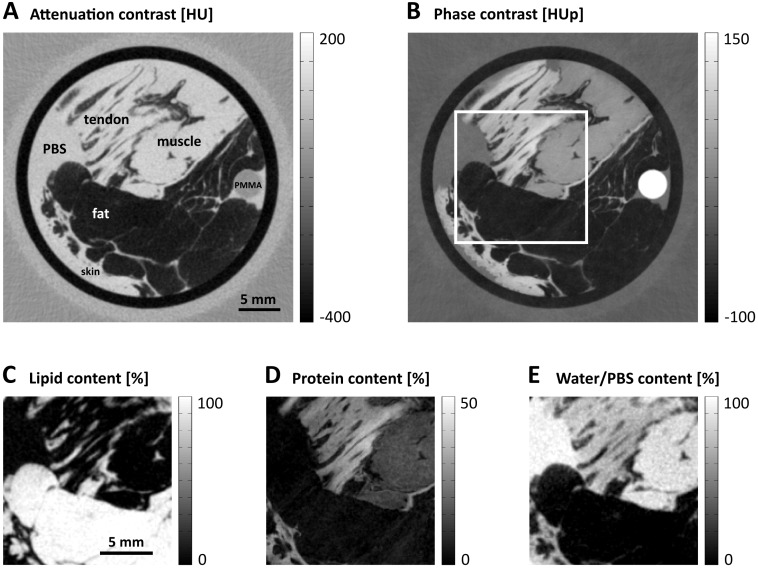
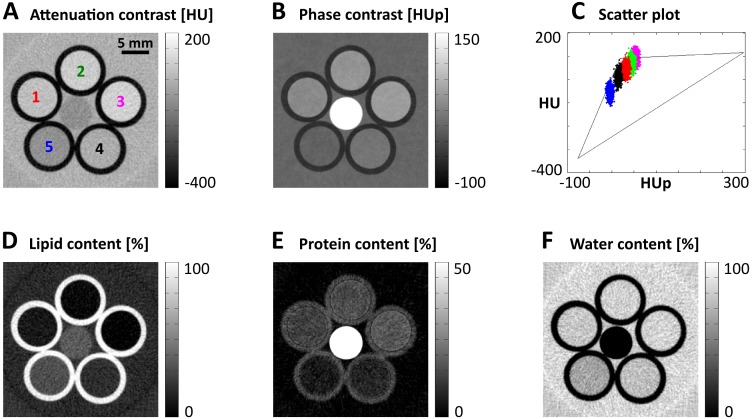
X-ray phase-contrast computed tomography is an emerging imaging technology with powerful capabilities for three-dimensional (3D) visualization of weakly absorbing objects such as biological soft tissues. This technique is an extension of existing X-ray applications because conventional attenuation-contrast images are simultaneously acquired. The complementary information provided by both the contrast modalities suggests that enhanced material characterization is possible when performing combined data analysis. In this study, we describe how protein, lipid, and water concentrations in each 3D voxel can be quantified by vector decomposition. Experimental results of dairy products, porcine fat and rind, and different human soft tissue types are presented. The results demonstrate the potential of phase-contrast imaging as a new analysis tool. The 3D representations of protein, lipid, and water contents open up new opportunities in the fields of biology, medicine, and food science.

摘要

X射线相衬计算机断层扫描是一种新兴的成像技术,在对生物软组织等弱吸收物体进行三维(3D)可视化方面具有强大的能力。由于同时获取了传统的衰减对比图像,该技术是现有X射线应用的扩展。两种对比模式提供的互补信息表明,在进行联合数据分析时,可以增强材料表征。在本研究中,我们描述了如何通过矢量分解对每个3D体素中的蛋白质、脂质和水浓度进行量化。给出了乳制品、猪脂肪和猪皮以及不同人类软组织类型的实验结果。结果证明了相衬成像作为一种新分析工具的潜力。蛋白质、脂质和水含量的3D表示在生物学、医学和食品科学领域开辟了新的机遇。

https://cdn.ncbi.nlm.nih.gov/pmc/blobs/8122/4803315/991775511586/pone.0151889.g004.jpg
https://cdn.ncbi.nlm.nih.gov/pmc/blobs/8122/4803315/a6f14d3bc1f6/pone.0151889.g001.jpg
https://cdn.ncbi.nlm.nih.gov/pmc/blobs/8122/4803315/392cef1ff4a2/pone.0151889.g002.jpg
https://cdn.ncbi.nlm.nih.gov/pmc/blobs/8122/4803315/ca58f420085d/pone.0151889.g003.jpg
https://cdn.ncbi.nlm.nih.gov/pmc/blobs/8122/4803315/991775511586/pone.0151889.g004.jpg

相似文献

1
Quantitative Three-Dimensional Imaging of Lipid, Protein, and Water Contents via X-Ray Phase-Contrast Tomography.通过X射线相衬断层扫描对脂质、蛋白质和水含量进行定量三维成像
PLoS One. 2016 Mar 22;11(3):e0151889. doi: 10.1371/journal.pone.0151889. eCollection 2016.
2
Novel X-ray phase-contrast tomography method for quantitative studies of heat induced structural changes in meat.新型 X 射线相衬断层摄影术定量研究热诱导肉结构变化。
Meat Sci. 2015 Feb;100:217-21. doi: 10.1016/j.meatsci.2014.10.009.
3
Theory and preliminary experimental verification of quantitative edge illumination x-ray phase contrast tomography.定量边缘照明X射线相衬断层扫描的理论与初步实验验证
Opt Express. 2014 Apr 7;22(7):7989-8000. doi: 10.1364/OE.22.007989.
4
A micro-tomography method based on X-ray diffraction enhanced imaging for the visualization of micro-organs and soft tissues.一种基于X射线衍射增强成像的显微断层扫描方法,用于微器官和软组织的可视化。
Comput Med Imaging Graph. 2006 Sep-Oct;30(6-7):339-47. doi: 10.1016/j.compmedimag.2006.09.003. Epub 2006 Oct 18.
5
X-ray phase contrast tomography by tracking near field speckle.通过跟踪近场散斑进行X射线相衬断层扫描
Sci Rep. 2015 Mar 4;5:8762. doi: 10.1038/srep08762.
6
Low-dose phase contrast tomography with conventional x-ray sources.使用传统X射线源的低剂量相衬断层扫描。
Med Phys. 2014 Jul;41(7):070701. doi: 10.1118/1.4884297.
7
Phase-Contrast Hounsfield Units of Fixated and Non-Fixated Soft-Tissue Samples.固定和未固定软组织样本的相位对比亨氏单位
PLoS One. 2015 Aug 31;10(8):e0137016. doi: 10.1371/journal.pone.0137016. eCollection 2015.
8
Microscopic dual-energy CT (microDECT): a flexible tool for multichannel ex vivo 3D imaging of biological specimens.微观双能CT(microDECT):用于生物标本多通道离体三维成像的灵活工具。
J Microsc. 2017 Jul;267(1):3-26. doi: 10.1111/jmi.12543. Epub 2017 Mar 7.
9
Implementation of dual- and triple-energy cone-beam micro-CT for postreconstruction material decomposition.双能和三能锥束显微CT在重建后材料分解中的应用。
Med Phys. 2008 Nov;35(11):5030-42. doi: 10.1118/1.2987668.
10
Feasibility Study of the 3D Visualization at High Resolution of Intra-Cranial Rabbit Eyes With X-Ray CT Phase-Contrast Imaging.X射线CT相衬成像对兔颅内眼进行高分辨率三维可视化的可行性研究
Invest Ophthalmol Vis Sci. 2017 Nov 1;58(13):5941-5948. doi: 10.1167/iovs.17-22273.

引用本文的文献

1
Quantification of water and lipid composition of perivascular adipose tissue using coronary CT angiography: a simulation study.使用冠状动脉CT血管造影术对血管周围脂肪组织的水和脂质成分进行定量分析:一项模拟研究。
Int J Cardiovasc Imaging. 2025 Jun;41(6):1091-1101. doi: 10.1007/s10554-025-03390-1. Epub 2025 Apr 10.
2
Exploring single-shot propagation and speckle based phase recovery techniques for object thickness estimation by using a polychromatic X-ray laboratory source.利用多色X射线实验室光源探索基于单次传播和散斑的相位恢复技术用于物体厚度估计。
J Med Imaging (Bellingham). 2024 Jul;11(4):043501. doi: 10.1117/1.JMI.11.4.043501. Epub 2024 Jul 25.
3

本文引用的文献

1
Complementary X-ray tomography techniques for histology-validated 3D imaging of soft and hard tissues using plaque-containing blood vessels as examples.使用含斑块的血管作为示例,用于对软组织和硬组织进行组织学验证的 3D 成像的补充 X 射线断层成像技术。
Nat Protoc. 2014;9(6):1401-15. doi: 10.1038/nprot.2014.091. Epub 2014 May 22.
2
Breast tissue characterization with photon-counting spectral CT imaging: a postmortem breast study.基于光子计数光谱CT成像的乳腺组织特征分析:一项乳腺尸体研究
Radiology. 2014 Sep;272(3):731-8. doi: 10.1148/radiol.14132732. Epub 2014 May 7.
3
Quantitative breast tissue characterization using grating-based x-ray phase-contrast imaging.
X-ray phase-contrast microtomography of soft tissues using a compact laboratory system with two-directional sensitivity.
使用具有双向灵敏度的紧凑型实验室系统对软组织进行X射线相衬显微断层成像。
Optica. 2023 Jun 30;10(7):880-887. doi: 10.1364/OPTICA.487270. eCollection 2023 Jul 20.
4
Hierarchical imaging and computational analysis of three-dimensional vascular network architecture in the entire postnatal and adult mouse brain.对整个新生期和成年期小鼠大脑中的三维血管网络结构进行层次成像和计算分析。
Nat Protoc. 2021 Oct;16(10):4564-4610. doi: 10.1038/s41596-021-00587-1. Epub 2021 Sep 3.
5
Quantitative X-ray phase contrast computed tomography with grating interferometry : Biomedical applications of quantitative X-ray grating-based phase contrast computed tomography.基于光栅的定量 X 射线相衬计算机断层成像术:定量 X 射线光栅相衬计算机断层成像术的生物医学应用。
Eur J Nucl Med Mol Imaging. 2021 Dec;48(13):4171-4188. doi: 10.1007/s00259-021-05259-6. Epub 2021 Apr 13.
6
3D grating-based X-ray phase-contrast computed tomography for high-resolution quantitative assessment of cartilage: An experimental feasibility study with 3T MRI, 7T MRI and biomechanical correlation.基于 3D 光栅的 X 射线相衬计算机断层扫描用于高分辨率定量评估软骨:3T MRI、7T MRI 和生物力学相关性的实验可行性研究。
PLoS One. 2019 Feb 14;14(2):e0212106. doi: 10.1371/journal.pone.0212106. eCollection 2019.
7
High resolution laboratory grating-based X-ray phase-contrast CT.高分辨率实验室光栅式 X 射线相衬 CT。
Sci Rep. 2018 Oct 26;8(1):15884. doi: 10.1038/s41598-018-33997-5.
8
Review of quantitative multiscale imaging of breast cancer.乳腺癌定量多尺度成像综述。
J Med Imaging (Bellingham). 2018 Jan;5(1):010901. doi: 10.1117/1.JMI.5.1.010901. Epub 2018 Jan 22.
使用基于光栅的X射线相衬成像进行乳腺组织定量表征。
Phys Med Biol. 2014 Apr 7;59(7):1557-71. doi: 10.1088/0031-9155/59/7/1557. Epub 2014 Mar 10.
4
In-vivo dark-field and phase-contrast x-ray imaging.体内暗场与相衬X射线成像。
Sci Rep. 2013 Nov 13;3:3209. doi: 10.1038/srep03209.
5
MR quantitative biomarkers of non-alcoholic fatty liver disease: technical evolutions and future trends.MR 定量生物标志物在非酒精性脂肪性肝病中的应用:技术进展与未来趋势。
Quant Imaging Med Surg. 2013 Aug;3(4):192-5. doi: 10.3978/j.issn.2223-4292.2013.08.01.
6
Effect of increasing the protein-to-fat ratio and reducing fat content on the chemical and physical properties of processed cheese product.提高蛋白质与脂肪比例及降低脂肪含量对再制奶酪产品化学和物理性质的影响。
J Dairy Sci. 2013;96(11):6830-6839. doi: 10.3168/jds.2013-6685. Epub 2013 Aug 29.
7
Quantitative X-ray phase-contrast computed tomography at 82 keV.82千电子伏特的定量X射线相衬计算机断层扫描
Opt Express. 2013 Feb 25;21(4):4155-66. doi: 10.1364/OE.21.004155.
8
X-ray phase-contrast imaging: from pre-clinical applications towards clinics.X 射线相衬成像:从临床前应用走向临床。
Phys Med Biol. 2013 Jan 7;58(1):R1-35. doi: 10.1088/0031-9155/58/1/R1. Epub 2012 Dec 10.
9
High-resolution, low-dose phase contrast X-ray tomography for 3D diagnosis of human breast cancers.高分辨率、低剂量的相衬 X 射线断层扫描术用于人类乳腺癌的 3D 诊断。
Proc Natl Acad Sci U S A. 2012 Nov 6;109(45):18290-4. doi: 10.1073/pnas.1204460109. Epub 2012 Oct 22.
10
Experimental results from a preclinical X-ray phase-contrast CT scanner.临床前 X 射线相衬 CT 扫描仪的实验结果。
Proc Natl Acad Sci U S A. 2012 Sep 25;109(39):15691-6. doi: 10.1073/pnas.1207503109. Epub 2012 Sep 10.