Lewandowska Marta K, Radivojević Miloš, Jäckel David, Müller Jan, Hierlemann Andreas R
Bio Engineering Laboratory, Department of Biosystems Science and Engineering, ETH Zürich Basel, Switzerland.
Front Neurosci. 2016 Mar 7;10:83. doi: 10.3389/fnins.2016.00083. eCollection 2016.
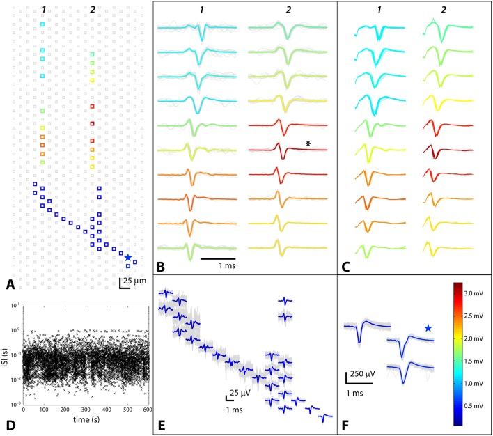
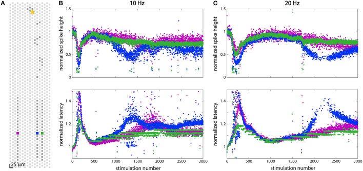
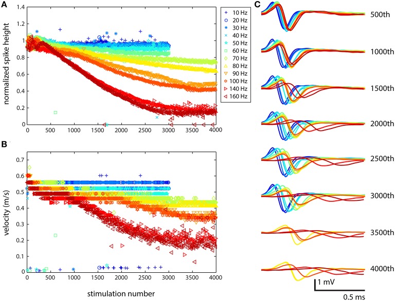
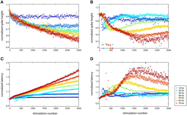
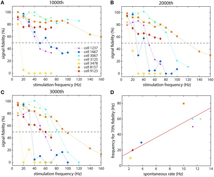
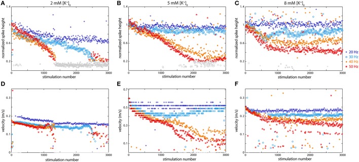
Mammalian cortical axons are extremely thin processes that are difficult to study as a result of their small diameter: they are too narrow to patch while intact, and super-resolution microscopy is needed to resolve single axons. We present a method for studying axonal physiology by pairing a high-density microelectrode array with a microfluidic axonal isolation device, and use it to study activity-dependent modulation of axonal signal propagation evoked by stimulation near the soma. Up to three axonal branches from a single neuron, isolated in different channels, were recorded from simultaneously using 10-20 electrodes per channel. The axonal channels amplified spikes such that propagations of individual signals along tens of electrodes could easily be discerned with high signal to noise. Stimulation from 10 up to 160 Hz demonstrated similar qualitative results from all of the cells studied: extracellular action potential characteristics changed drastically in response to stimulation. Spike height decreased, spike width increased, and latency increased, as a result of reduced propagation velocity, as the number of stimulations and the stimulation frequencies increased. Quantitatively, the strength of these changes manifested itself differently in cells at different frequencies of stimulation. Some cells' signal fidelity fell to 80% already at 10 Hz, while others maintained 80% signal fidelity at 80 Hz. Differences in modulation by axonal branches of the same cell were also seen for different stimulation frequencies, starting at 10 Hz. Potassium ion concentration changes altered the behavior of the cells causing propagation failures at lower concentrations and improving signal fidelity at higher concentrations.
哺乳动物的皮质轴突是极其纤细的突起,由于其直径小而难以研究:它们太细,完整时无法进行膜片钳操作,需要超分辨率显微镜才能分辨单个轴突。我们提出了一种将高密度微电极阵列与微流控轴突分离装置相结合来研究轴突生理学的方法,并利用该方法研究由胞体附近刺激诱发的轴突信号传播的活动依赖性调制。在不同通道中分离出的单个神经元的多达三个轴突分支,每个通道使用10 - 20个电极同时进行记录。轴突通道放大了尖峰信号,使得单个信号沿数十个电极的传播能够以高信噪比轻松辨别。从10 Hz到160 Hz的刺激对所有研究的细胞都显示出相似的定性结果:细胞外动作电位特征因刺激而发生显著变化。随着刺激次数和刺激频率的增加,由于传播速度降低,尖峰高度降低,尖峰宽度增加,潜伏期延长。在数量上,这些变化的强度在不同刺激频率的细胞中表现不同。一些细胞在10 Hz时信号保真度就已降至80%,而另一些细胞在80 Hz时仍保持80%的信号保真度。从10 Hz开始,在不同刺激频率下,同一细胞的轴突分支的调制差异也很明显。钾离子浓度变化改变了细胞的行为,在较低浓度下导致传播失败,在较高浓度下提高信号保真度。