• 文献检索
  • 文档翻译
  • 深度研究
  • 学术资讯
  • Suppr Zotero 插件Zotero 插件
  • 邀请有礼
  • 套餐&价格
  • 历史记录
应用&插件
Suppr Zotero 插件Zotero 插件浏览器插件Mac 客户端Windows 客户端微信小程序
定价
高级版会员购买积分包购买API积分包
服务
文献检索文档翻译深度研究API 文档MCP 服务
关于我们
关于 Suppr公司介绍联系我们用户协议隐私条款
关注我们

Suppr 超能文献

核心技术专利:CN118964589B侵权必究
粤ICP备2023148730 号-1Suppr @ 2026

文献检索

告别复杂PubMed语法,用中文像聊天一样搜索,搜遍4000万医学文献。AI智能推荐,让科研检索更轻松。

立即免费搜索

文件翻译

保留排版,准确专业,支持PDF/Word/PPT等文件格式,支持 12+语言互译。

免费翻译文档

深度研究

AI帮你快速写综述,25分钟生成高质量综述,智能提取关键信息,辅助科研写作。

立即免费体验

揭示神经元树突上的跨膜电流沿树突分布。

Revealing the distribution of transmembrane currents along the dendritic tree of a neuron from extracellular recordings.

机构信息

Wigner Research Centre for Physics, Hungarian Academy of Sciences, Budapest, Hungary.

Institute of Cognitive Neuroscience and Psychology, Research Centre for Natural Sciences, Hungarian Academy of Sciences, Budapest, Hungary.

出版信息

Elife. 2017 Nov 17;6:e29384. doi: 10.7554/eLife.29384.

DOI:10.7554/eLife.29384
PMID:29148974
原文链接:https://pmc.ncbi.nlm.nih.gov/articles/PMC5716668/
Abstract

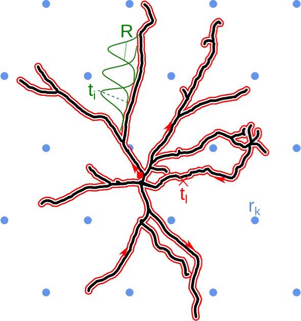
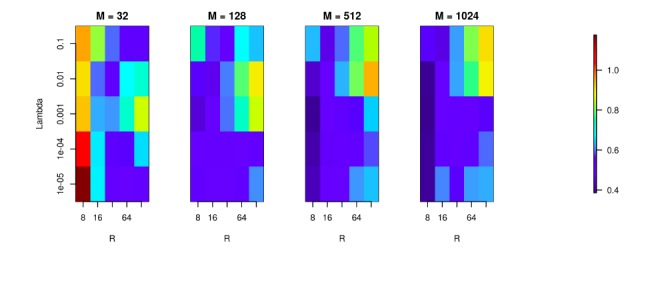
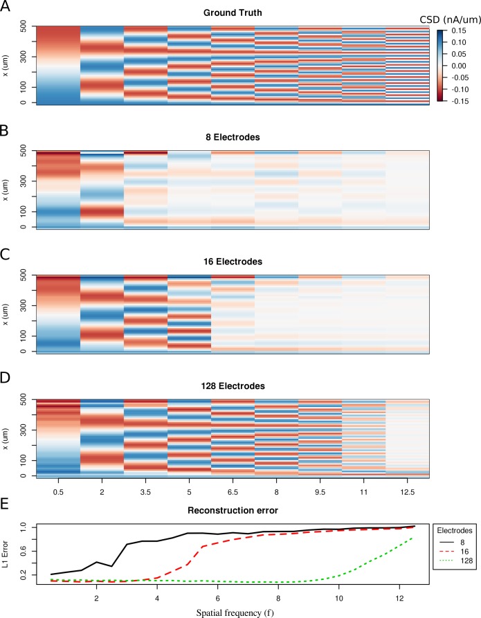
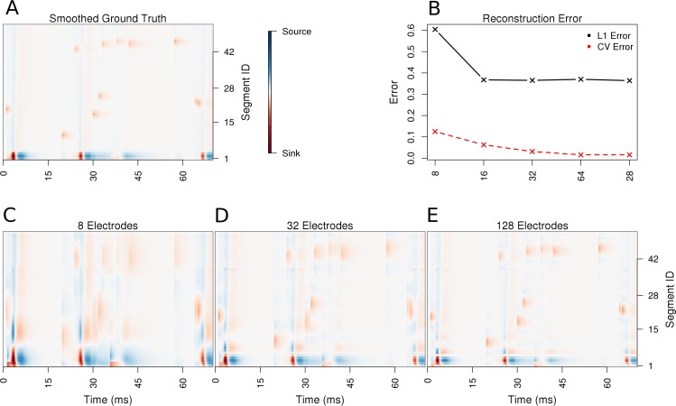
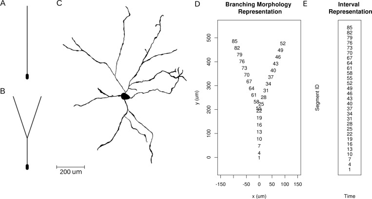
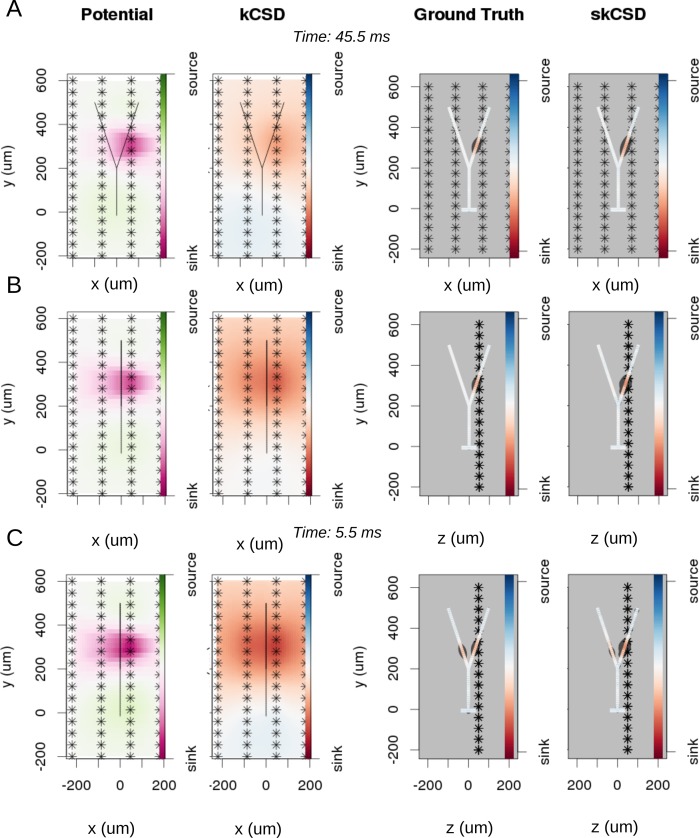
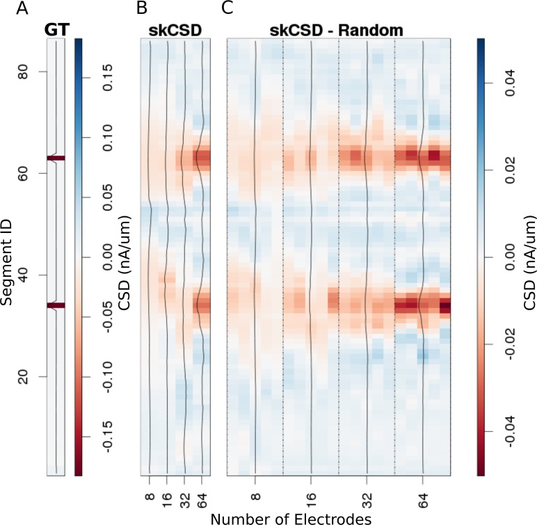
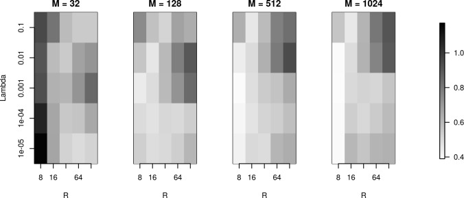
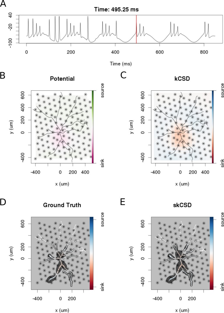
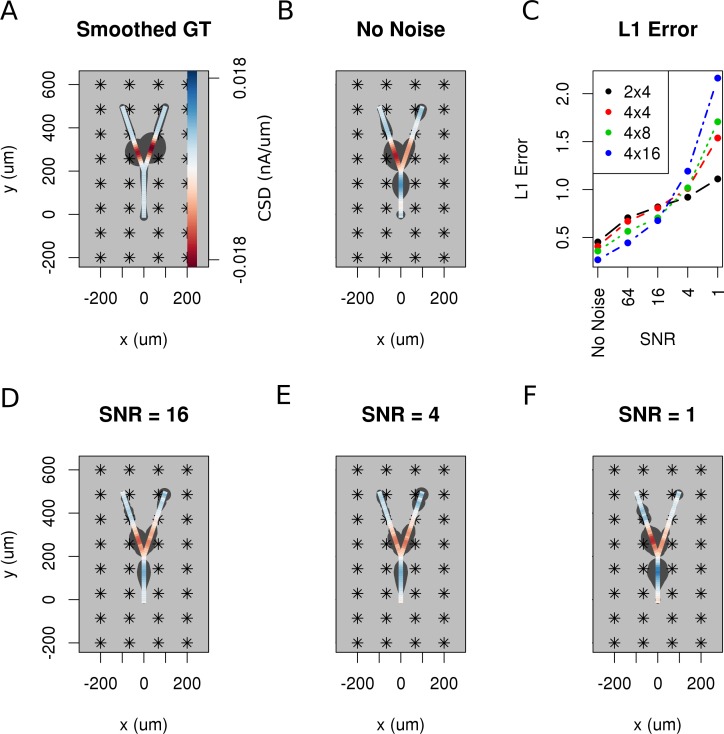
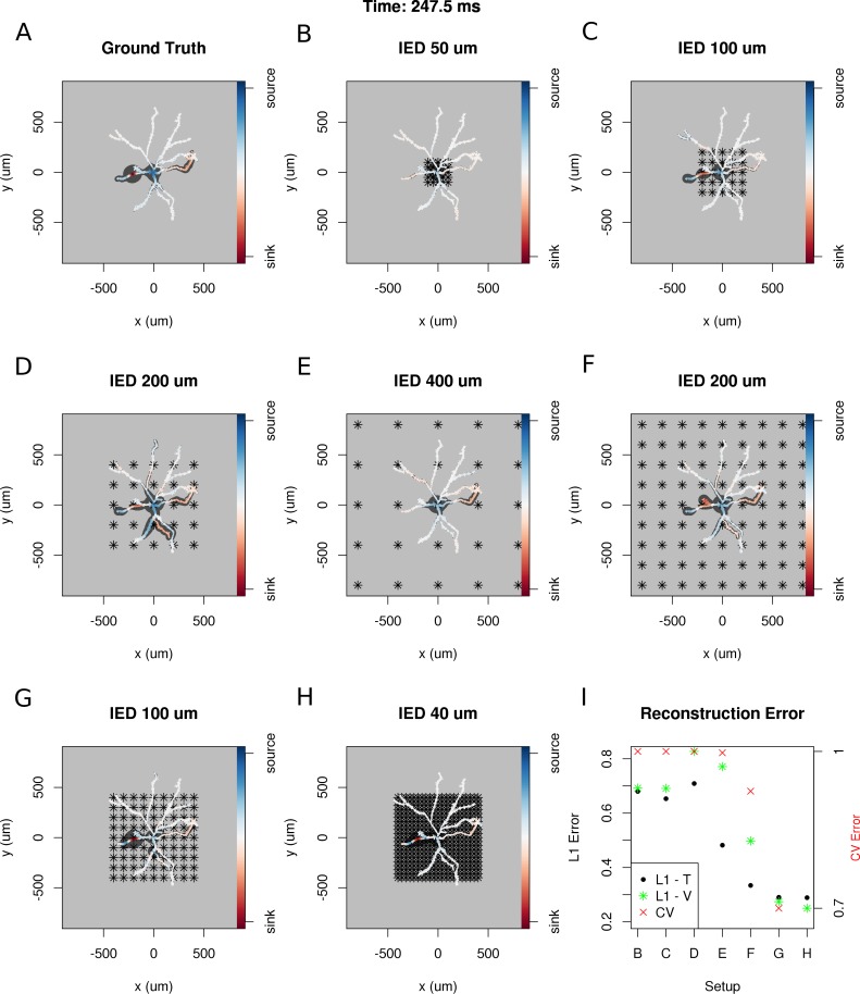
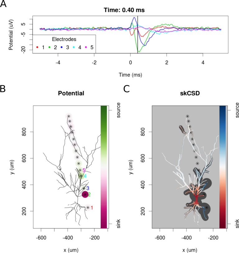
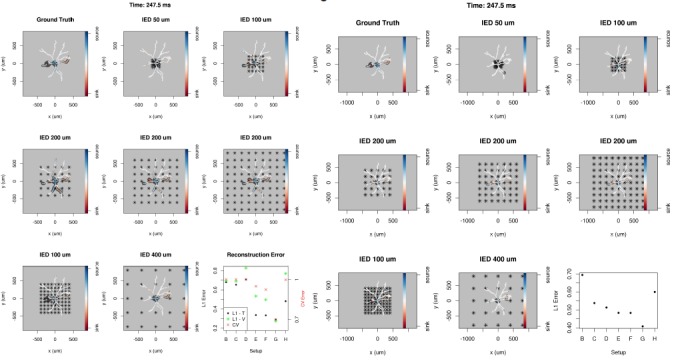
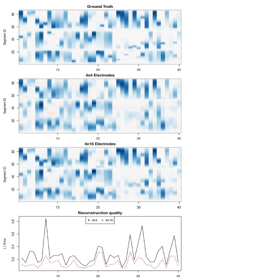
Revealing the current source distribution along the neuronal membrane is a key step on the way to understanding neural computations; however, the experimental and theoretical tools to achieve sufficient spatiotemporal resolution for the estimation remain to be established. Here, we address this problem using extracellularly recorded potentials with arbitrarily distributed electrodes for a neuron of known morphology. We use simulations of models with varying complexity to validate the proposed method and to give recommendations for experimental applications. The method is applied to in vitro data from rat hippocampus.

摘要

揭示神经元膜上的电流源分布是理解神经计算的关键步骤;然而,实现足够的时空分辨率进行估计的实验和理论工具仍有待建立。在这里,我们使用具有任意分布电极的细胞外记录电势来解决这个问题,这些电极用于已知形态的神经元。我们使用具有不同复杂度的模型的模拟来验证所提出的方法,并为实验应用提供建议。该方法应用于大鼠海马体的体外数据。

https://cdn.ncbi.nlm.nih.gov/pmc/blobs/8ba6/5716668/b35bf7cf95bf/elife-29384-resp-fig3.jpg
https://cdn.ncbi.nlm.nih.gov/pmc/blobs/8ba6/5716668/52b51003d1f0/elife-29384-fig1.jpg
https://cdn.ncbi.nlm.nih.gov/pmc/blobs/8ba6/5716668/a7500c6a09ac/elife-29384-fig2.jpg
https://cdn.ncbi.nlm.nih.gov/pmc/blobs/8ba6/5716668/d6ed3b7b76be/elife-29384-fig3.jpg
https://cdn.ncbi.nlm.nih.gov/pmc/blobs/8ba6/5716668/12089b1ba035/elife-29384-fig4.jpg
https://cdn.ncbi.nlm.nih.gov/pmc/blobs/8ba6/5716668/8357a90ab82d/elife-29384-fig5.jpg
https://cdn.ncbi.nlm.nih.gov/pmc/blobs/8ba6/5716668/8f43192ed1d2/elife-29384-fig6.jpg
https://cdn.ncbi.nlm.nih.gov/pmc/blobs/8ba6/5716668/3211b8d492a8/elife-29384-fig7.jpg
https://cdn.ncbi.nlm.nih.gov/pmc/blobs/8ba6/5716668/49f58d605028/elife-29384-fig8.jpg
https://cdn.ncbi.nlm.nih.gov/pmc/blobs/8ba6/5716668/833c29d78653/elife-29384-fig9.jpg
https://cdn.ncbi.nlm.nih.gov/pmc/blobs/8ba6/5716668/fa0c58097611/elife-29384-fig10.jpg
https://cdn.ncbi.nlm.nih.gov/pmc/blobs/8ba6/5716668/287e5610884d/elife-29384-fig11.jpg
https://cdn.ncbi.nlm.nih.gov/pmc/blobs/8ba6/5716668/2cb537cb13f0/elife-29384-resp-fig1.jpg
https://cdn.ncbi.nlm.nih.gov/pmc/blobs/8ba6/5716668/c04519f3c3fd/elife-29384-resp-fig2.jpg
https://cdn.ncbi.nlm.nih.gov/pmc/blobs/8ba6/5716668/b35bf7cf95bf/elife-29384-resp-fig3.jpg

相似文献

1
Revealing the distribution of transmembrane currents along the dendritic tree of a neuron from extracellular recordings.揭示神经元树突上的跨膜电流沿树突分布。
Elife. 2017 Nov 17;6:e29384. doi: 10.7554/eLife.29384.
2
Seeing beyond the spikes: reconstructing the complete spatiotemporal membrane potential distribution from paired intra- and extracellular recordings.透过尖峰信号洞察本质:从细胞内和细胞外配对记录重建完整的时空膜电位分布
J Physiol. 2023 Aug;601(15):3351-3376. doi: 10.1113/JP283550. Epub 2023 Jan 25.
3
Kernel current source density method.内核电流源密度方法。
Neural Comput. 2012 Feb;24(2):541-75. doi: 10.1162/NECO_a_00236. Epub 2011 Nov 17.
4
waveCSD: A method for estimating transmembrane currents originated from propagating neuronal activity in the neocortex: Application to study cortical spreading depression.波 CSD:一种在新皮层中估算源自传播性神经元活动的跨膜电流的方法:在研究皮质扩散性抑制中的应用。
J Neurosci Methods. 2018 Sep 1;307:106-124. doi: 10.1016/j.jneumeth.2018.06.024. Epub 2018 Jul 3.
5
Ion diffusion may introduce spurious current sources in current-source density (CSD) analysis.离子扩散可能在电流源密度(CSD)分析中引入虚假电流源。
J Neurophysiol. 2017 Jul 1;118(1):114-120. doi: 10.1152/jn.00976.2016. Epub 2017 Mar 15.
6
Distribution and functional properties of 5-HT3 receptors in the rat hippocampal dentate gyrus: a patch-clamp study.
J Neurophysiol. 1994 May;71(5):1935-47. doi: 10.1152/jn.1994.71.5.1935.
7
Space-clamp problems when voltage clamping neurons expressing voltage-gated conductances.对表达电压门控电导的神经元进行电压钳制时的空间钳制问题。
J Neurophysiol. 2008 Mar;99(3):1127-36. doi: 10.1152/jn.01232.2007. Epub 2008 Jan 9.
8
Electrotonic profiles of interneurons in stratum pyramidale of the CA1 region of rat hippocampus.大鼠海马体CA1区锥体层中间神经元的电紧张特性
J Neurophysiol. 1994 May;71(5):1948-58. doi: 10.1152/jn.1994.71.5.1948.
9
Contributions of space-clamp errors to apparent time-dependent loss of Mg block induced by NMDA.空间钳制误差对NMDA诱导的Mg2+阻断明显的时间依赖性丧失的作用。
J Neurophysiol. 2017 Jul 1;118(1):532-543. doi: 10.1152/jn.00106.2017. Epub 2017 Mar 29.
10
A model for dendritic Ca2+ accumulation in hippocampal pyramidal neurons based on fluorescence imaging measurements.基于荧光成像测量的海马锥体神经元树突状Ca2+积累模型。
J Neurophysiol. 1994 Mar;71(3):1065-77. doi: 10.1152/jn.1994.71.3.1065.

引用本文的文献

1
Advanced neuroprosthetic electrode design optimized by electromagnetic finite element simulation: innovations and applications.通过电磁有限元模拟优化的先进神经假体电极设计:创新与应用
Front Bioeng Biotechnol. 2024 Nov 6;12:1476447. doi: 10.3389/fbioe.2024.1476447. eCollection 2024.
2
kCSD-python, reliable current source density estimation with quality control.kCSD-python,具有质量控制的可靠电流源密度估计。
PLoS Comput Biol. 2024 Mar 14;20(3):e1011941. doi: 10.1371/journal.pcbi.1011941. eCollection 2024 Mar.
3
Computing Extracellular Electric Potentials from Neuronal Simulations.

本文引用的文献

1
Combination of High-density Microelectrode Array and Patch Clamp Recordings to Enable Studies of Multisynaptic Integration.高密度微电极阵列与膜片钳记录的联合应用实现了对多突触整合的研究。
Sci Rep. 2017 Apr 20;7(1):978. doi: 10.1038/s41598-017-00981-4.
2
Collection of Simulated Data from a Thalamocortical Network Model.从丘脑皮质网络模型收集模拟数据。
Neuroinformatics. 2017 Jan;15(1):87-99. doi: 10.1007/s12021-016-9319-4.
3
Validating silicon polytrodes with paired juxtacellular recordings: method and dataset.使用配对细胞旁记录法验证硅多电极:方法与数据集
从神经元模拟中计算细胞外电势。
Adv Exp Med Biol. 2022;1359:179-199. doi: 10.1007/978-3-030-89439-9_8.
4
What we can and what we cannot see with extracellular multielectrodes.细胞外多电极能看到什么和不能看到什么。
PLoS Comput Biol. 2021 May 14;17(5):e1008615. doi: 10.1371/journal.pcbi.1008615. eCollection 2021 May.
5
Application of a flexible polymer microECoG array to map functional coherence in schizophrenia model.柔性聚合物微脑电图阵列在精神分裂症模型中映射功能连贯性的应用。
MethodsX. 2020 Oct 22;7:101117. doi: 10.1016/j.mex.2020.101117. eCollection 2020.
6
Multimodal Modeling of Neural Network Activity: Computing LFP, ECoG, EEG, and MEG Signals With LFPy 2.0.神经网络活动的多模态建模:使用LFPy 2.0计算局部场电位、皮层脑电图、脑电图和脑磁图信号
Front Neuroinform. 2018 Dec 18;12:92. doi: 10.3389/fninf.2018.00092. eCollection 2018.
J Neurophysiol. 2016 Aug 1;116(2):892-903. doi: 10.1152/jn.00103.2016. Epub 2016 Jun 15.
4
Cortical Axons, Isolated in Channels, Display Activity-Dependent Signal Modulation as a Result of Targeted Stimulation.在通道中分离出的皮质轴突,由于靶向刺激而显示出活动依赖性信号调制。
Front Neurosci. 2016 Mar 7;10:83. doi: 10.3389/fnins.2016.00083. eCollection 2016.
5
Generalized Laminar Population Analysis (gLPA) for Interpretation of Multielectrode Data from Cortex.用于解释来自皮质的多电极数据的广义层状群体分析(gLPA)
Front Neuroinform. 2016 Jan 25;10:1. doi: 10.3389/fninf.2016.00001. eCollection 2016.
6
Spike Detection for Large Neural Populations Using High Density Multielectrode Arrays.使用高密度多电极阵列对大型神经群体进行尖峰检测。
Front Neuroinform. 2015 Dec 18;9:28. doi: 10.3389/fninf.2015.00028. eCollection 2015.
7
Combined two-photon imaging, electrophysiological, and anatomical investigation of the human neocortex in vitro.体外人类新皮层的双光子成像、电生理和解剖学联合研究。
Neurophotonics. 2014 Jul;1(1):011013. doi: 10.1117/1.NPh.1.1.011013. Epub 2014 Sep 11.
8
Past, present and future of spike sorting techniques.尖峰分类技术的过去、现在与未来。
Brain Res Bull. 2015 Oct;119(Pt B):106-17. doi: 10.1016/j.brainresbull.2015.04.007. Epub 2015 Apr 27.
9
Modelling and Analysis of Electrical Potentials Recorded in Microelectrode Arrays (MEAs).微电极阵列(MEA)记录的电势建模与分析
Neuroinformatics. 2015 Oct;13(4):403-26. doi: 10.1007/s12021-015-9265-6.
10
Revealing neuronal function through microelectrode array recordings.通过微电极阵列记录揭示神经元功能。
Front Neurosci. 2015 Jan 6;8:423. doi: 10.3389/fnins.2014.00423. eCollection 2014.