van der Meijden K, Bravenboer N, Dirks N F, Heijboer A C, den Heijer M, de Wit G M J, Offringa C, Lips P, Jaspers R T
Department of Internal Medicine/Endocrinology, VU University Medical Center, MOVE Research Institute Amsterdam, Amsterdam, The Netherlands.
Department of Clinical Chemistry, VU University Medical Center, MOVE Research Institute Amsterdam, Amsterdam, The Netherlands.
J Cell Physiol. 2016 Nov;231(11):2517-28. doi: 10.1002/jcp.25388. Epub 2016 Apr 14.
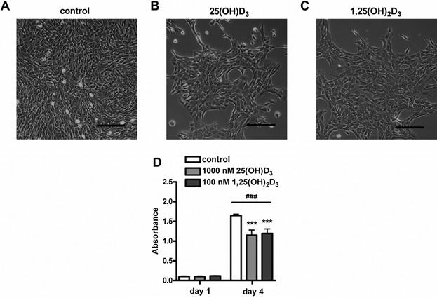
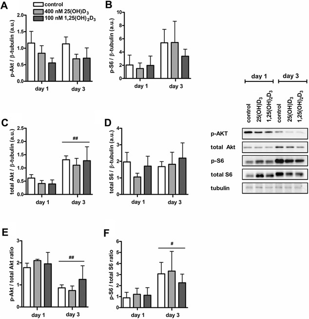
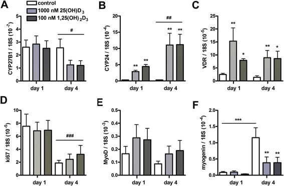
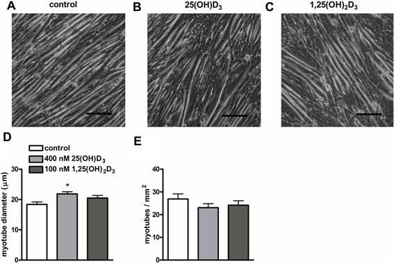
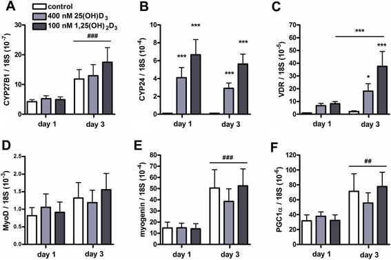
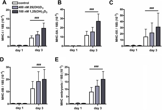
An adequate vitamin D status is essential to optimize muscle strength. However, whether vitamin D directly reduces muscle fiber atrophy or stimulates muscle fiber hypertrophy remains subject of debate. A mechanism that may affect the role of vitamin D in the regulation of muscle fiber size is the local conversion of 25(OH)D to 1,25(OH)2 D by 1α-hydroxylase. Therefore, we investigated in a murine C2C12 myoblast culture whether both 1,25(OH)2 D3 and 25(OH)D3 affect myoblast proliferation, differentiation, and myotube size and whether these cells are able to metabolize 25(OH)D3 and 1,25(OH)2 D3 . We show that myoblasts not only responded to 1,25(OH)2 D3 , but also to the precursor 25(OH)D3 by increasing their VDR mRNA expression and reducing their proliferation. In differentiating myoblasts and myotubes 1,25(OH)2 D3 as well as 25(OH)D3 stimulated VDR mRNA expression and in myotubes 1,25(OH)2 D3 also stimulated MHC mRNA expression. However, this occurred without notable effects on myotube size. Moreover, no effects on the Akt/mTOR signaling pathway as well as MyoD and myogenin mRNA levels were observed. Interestingly, both myoblasts and myotubes expressed CYP27B1 and CYP24 mRNA which are required for vitamin D3 metabolism. Although 1α-hydroxylase activity could not be shown in myotubes, after treatment with 1,25(OH)2 D3 or 25(OH)D3 myotubes showed strongly elevated CYP24 mRNA levels compared to untreated cells. Moreover, myotubes were able to convert 25(OH)D3 to 24R,25(OH)2 D3 which may play a role in myoblast proliferation and differentiation. These data suggest that skeletal muscle is not only a direct target for vitamin D3 metabolites, but is also able to metabolize 25(OH)D3 and 1,25(OH)2 D3 . J. Cell. Physiol. 231: 2517-2528, 2016. © 2016 The Authors. Journal of Cellular Physiology Published by Wiley Periodicals, Inc.
充足的维生素D水平对于优化肌肉力量至关重要。然而,维生素D是直接减少肌纤维萎缩还是刺激肌纤维肥大仍存在争议。1α-羟化酶将25(OH)D局部转化为1,25(OH)2D这一机制可能会影响维生素D在调节肌纤维大小中的作用。因此,我们在小鼠C2C12成肌细胞培养中研究了1,25(OH)2D3和25(OH)D3是否会影响成肌细胞的增殖、分化以及肌管大小,以及这些细胞是否能够代谢25(OH)D3和1,25(OH)2D3。我们发现,成肌细胞不仅对1,25(OH)2D3有反应,对前体25(OH)D3也有反应,表现为其VDR mRNA表达增加且增殖减少。在分化的成肌细胞和肌管中,1,25(OH)2D3以及25(OH)D3均刺激VDR mRNA表达,在肌管中1,25(OH)2D3还刺激MHC mRNA表达。然而,这并未对肌管大小产生显著影响。此外,未观察到对Akt/mTOR信号通路以及MyoD和肌细胞生成素mRNA水平有影响。有趣的是,成肌细胞和肌管均表达维生素D3代谢所需的CYP27B1和CYP24 mRNA。虽然在肌管中未显示出1α-羟化酶活性,但在用1,25(OH)2D3或25(OH)D3处理后,与未处理细胞相比,肌管中CYP24 mRNA水平显著升高。此外,肌管能够将25(OH)D3转化为24R,25(OH)2D3,这可能在成肌细胞增殖和分化中发挥作用。这些数据表明,骨骼肌不仅是维生素D3代谢产物的直接靶标,还能够代谢25(OH)D3和1,25(OH)2D3。《细胞生理学杂志》2016年第231卷:2517 - 2528页。© 2016作者。《细胞生理学杂志》由威利期刊公司出版。