Min Hyunjung, Jang Yong Ho, Cho Ik-Hyun, Yu Seong-Woon, Lee Sung Joong
Department of Neuroscience and Dental Research Institute, School of Dentistry, Seoul National University, 1 Gwanak-ro, Gwanak-gu, Seoul, 08826, Republic of Korea.
Department of Convergence Medical Science, College of Oriental Medicine, Kyung Hee University, Seoul, 02447, Korea.
Mol Brain. 2016 Apr 19;9:42. doi: 10.1186/s13041-016-0225-3.
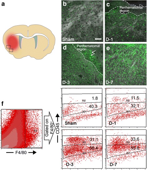
Intracerebral hemorrhage (ICH) is one of the major causes of stroke. After onset of ICH, massive infiltration of macrophages is detected in the peri-hematoma regions. Still, the function of these macrophages in ICH has not been completely elucidated.
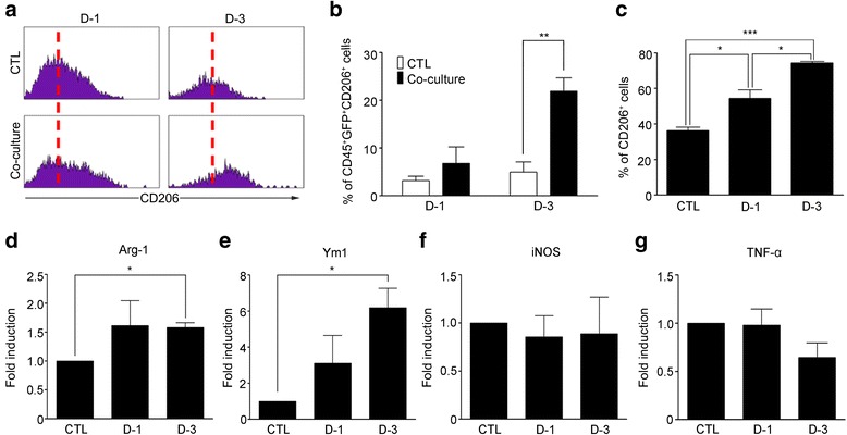
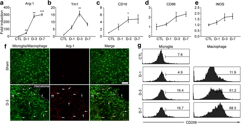
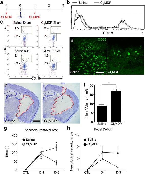
In a collagenase-induced ICH model, CX3CR1(+) macrophages accumulated in the peri-hematoma region. Characterization of these macrophages revealed expression of alternatively activated (M2) macrophage markers. In the macrophage-depleted mice, ICH-induced brain lesion volume was larger and neurological deficits were more severe compared to those of control mice, indicating a protective role of these macrophages in ICH. In the ICH-injured brain, mannose receptor-expressing macrophages increased at a delayed time point after ICH, indicating M2 polarization of the brain-infiltrating macrophages in the brain microenvironment. To explore this possibility, bone marrow-derived macrophages (BMDM) were co-cultured with mouse brain glial cells and then tested for activation phenotype. Upon co-culture with glia, the number of mannose receptor-positive M2 macrophages was significantly increased. Furthermore, treatment with glia-conditioned media increased the number of BMDM of M2 phenotype.
In this study, our data suggest that brain-infiltrating macrophages after ICH are polarized to the M2 phenotype by brain glial cells and thereby contribute to recovery from ICH injury.
脑出血(ICH)是中风的主要原因之一。脑出血发病后,在血肿周围区域可检测到大量巨噬细胞浸润。然而,这些巨噬细胞在脑出血中的功能尚未完全阐明。
在胶原酶诱导的脑出血模型中,CX3CR1(+)巨噬细胞在血肿周围区域积聚。对这些巨噬细胞的特征分析显示其表达替代性激活(M2)巨噬细胞标志物。在巨噬细胞耗竭的小鼠中,与对照小鼠相比,脑出血诱导的脑损伤体积更大,神经功能缺损更严重,表明这些巨噬细胞在脑出血中具有保护作用。在脑出血损伤的大脑中,表达甘露糖受体的巨噬细胞在脑出血后的延迟时间点增加,表明脑浸润巨噬细胞在脑微环境中发生M2极化。为了探究这种可能性,将骨髓来源的巨噬细胞(BMDM)与小鼠脑胶质细胞共培养,然后检测其激活表型。与胶质细胞共培养后,甘露糖受体阳性的M2巨噬细胞数量显著增加。此外,用胶质细胞条件培养基处理可增加M2表型BMDM的数量。
在本研究中,我们的数据表明,脑出血后脑浸润巨噬细胞被脑胶质细胞极化为M2表型,从而有助于从脑出血损伤中恢复。